Review
. 2018 Jan 2;128(1):85-96.
doi: 10.1172/JCI93562. Epub 2018 Jan 2.

The balancing act of the liver: tissue regeneration versus fibrosis

Review

The balancing act of the liver: tissue regeneration versus fibrosis

Lucía Cordero-Espinoza et al. J Clin Invest. .

Abstract

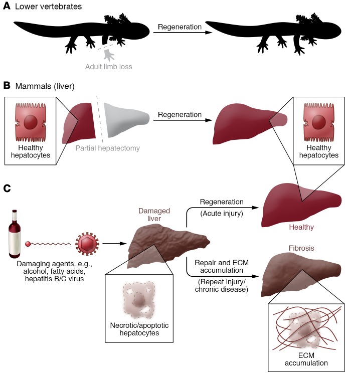
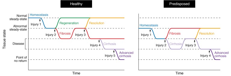
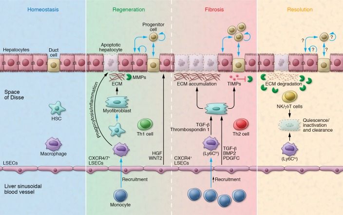
Epithelial cell loss alters a tissue's optimal function and awakens evolutionarily adapted healing mechanisms to reestablish homeostasis. Although adult mammalian organs have a limited regeneration potential, the liver stands out as one remarkable exception. Following injury, the liver mounts a dynamic multicellular response wherein stromal cells are activated in situ and/or recruited from the bloodstream, the extracellular matrix (ECM) is remodeled, and epithelial cells expand to replenish their lost numbers. Chronic damage makes this response persistent instead of transient, tipping the system into an abnormal steady state known as fibrosis, in which ECM accumulates excessively and tissue function degenerates. Here we explore the cellular and molecular switches that balance hepatic regeneration and fibrosis, with a focus on uncovering avenues of disease modeling and therapeutic intervention.


Conflict of interest statement

Conflict of interest: The authors have declared that no conflict of interest exists.

Figures

Figure 1
Figure 1. Coping with injury: regeneration versus repair.
(A) Lower vertebrates, such as axolotls, salamanders, and fish, are able to regenerate severed limbs through a process that reconstitutes original tissue anatomy and function without leaving a scar (a meshwork of ECM). Mammals may similarly regenerate complex tissues during embryogenesis, but lose most of this capacity in adulthood. (B) The liver is one of the few adult mammalian organs that retains a remarkable ability to regenerate itself. Resection of up to 70% of the liver mass via partial hepatectomy leads to compensatory growth from the intact tissue and fully restores organ size in a matter of days, similarly to axolotl limb regrowth. However, the hepatectomized liver is typically not injured or “damaged,” and regeneration is a result of the organ’s ability to sense insufficient size. (C) The liver may also regenerate following injury by exogenous and/or endogenous agents (e.g., alcohol, hepatitis B/C viruses, fatty acids) that cause hepatocyte death. This process is characterized by an inflammatory reaction and ECM synthesis/remodeling. However, if the damaging insult persists, the tissue will be repaired instead of regenerated, resulting in excessive scarring, known as fibrosis, that alters histoarchitecture and hinders optimal tissue function.
Figure 2
Figure 2. Periodicity of damage alters the ability of the tissue to return to homeostasis.
(Left) In healthy individuals, a punctual tissue injury (injury 1) to the liver awakens a regenerative response (green curve) to reestablish homeostasis or steady-state. Repeated injuries (injuries 1 + 2) hinder regeneration and make the system drift into a diseased state known as fibrosis (red curve). The tissue may recover from this as time progresses if no further damage is applied (resolution, injuries 1 + 2 + time, yellow curve). Alternatively, fibrosis will be maintained in the face of new damage (injuries 1+2+3, red curve). Additional injuries deteriorate the tissue until it reaches a cirrhotic (1 + 2 + 3 + 4, light purple curve) or advanced cirrhotic (1 + 2 + 3 + 4 + 5, dark purple curve) state. Recovery from this latter scenario is very unlikely. (Right) The tissue of predisposed individuals (e.g., aged) functions at an abnormal steady-state that makes them prone to develop fibrosis, thus accelerating disease progression and reaching a point of no recovery earlier.
Figure 3
Figure 3. Distinct cellular landscapes characterize homeostasis, regeneration, fibrosis, and resolution in the liver.
The homeostatic liver is characterized by rare cell proliferation and lack of de novo ECM deposition. During regeneration, epithelial replacement occurs predominantly via hepatocyte proliferation and, to a minor degree, through the activation of ductal progenitors. Resident (Kupffer) and bone marrow–recruited macrophages phagocytose the dead epithelium and launch an inflammatory cascade (e.g., TNF-α, IL-6). CXCR7/CXCR4+ LSECs provide mitogenic signals (HGF, WNT2) that sustain hepatocyte proliferation. HSCs transdifferentiate into myofibroblasts that deposit ECM on the wound site, although this matrix can be degraded via MMPs. In fibrosis, the hepatocyte compartment is highly senescent and ductal progenitor expansion becomes predominant. Monocyte-derived Ly6Chi macrophages (secreting TGF-β, thrombospondin 1) and CXCR4+ LSECs (secreting TGF-β, BMP2, and PDGFC) collectively enhance myofibroblast proliferation and survival. Myofibroblasts, in turn, secrete high levels of TIMPs, which inhibit MMPs and cause excessive matrix accumulation. A Th1- versus Th2-skewed immune system favors regeneration versus fibrosis, respectively. The resolution of fibrosis entails the return to quiescence/inactivation of myofibroblasts as well as their clearance by NK cells, γδ T cells, and Ly6Clo macrophages. High levels of MMPs contribute to matrix degradation. The mechanisms of epithelial replacement at this stage have not been fully elucidated.

References

    1. Gurtner G, Werner S, Barrandon Y, Longaker M. Wound repair and regeneration. Nature. 2008;453(7193):314–321. doi: 10.1038/nature07039. - DOI - PubMed
    1. Sánchez Alvarado A. Regeneration in the metazoans: why does it happen? Bioessays. 2000;22(6):578–590. doi: 10.1002/(SICI)1521-1878(200006)22:6<578::AID-BIES11>3.0.CO;2-#. - DOI - PubMed
    1. Timchenko NA. Aging and liver regeneration. Trends Endocrinol Metab. 2009;20(4):171–176. doi: 10.1016/j.tem.2009.01.005. - DOI - PubMed
    1. Michalopoulos GK. Liver regeneration. J Cell Physiol. 2007;213(2):286–300. doi: 10.1002/jcp.21172. - DOI - PMC - PubMed
    1. Taub R. Liver regeneration: from myth to mechanism. Nat Rev Mol Cell Biol. 2004;5(10):836–847. doi: 10.1038/nrm1489. - DOI - PubMed

Publication types