

Renal Tubular Cell Mitochondrial Dysfunction Occurs Despite Preserved Renal Oxygen Delivery in Experimental Septic Acute Kidney Injury
- PMID: 29293148
- PMCID: PMC5856355
- DOI: 10.1097/CCM.0000000000002937
Renal Tubular Cell Mitochondrial Dysfunction Occurs Despite Preserved Renal Oxygen Delivery in Experimental Septic Acute Kidney Injury
Abstract
Objective: To explain the paradigm of significant renal functional impairment despite preserved hemodynamics and histology in sepsis-induced acute kidney injury.
Design: Prospective observational animal study.
Setting: University research laboratory.
Subjects: Male Wistar rats.
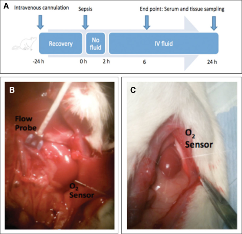
Intervention: Using a fluid-resuscitated sublethal rat model of fecal peritonitis, changes in renal function were characterized in relation to global and renal hemodynamics, and histology at 6 and 24 hours (n = 6-10). Sham-operated animals were used as comparison (n = 8). Tubular cell mitochondrial function was assessed using multiphoton confocal imaging of live kidney slices incubated in septic serum.
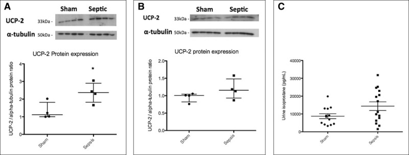
Measurements and main results: By 24 hours, serum creatinine was significantly elevated with a concurrent decrease in renal lactate clearance in septic animals compared with sham-operated and 6-hour septic animals. Renal uncoupling protein-2 was elevated in septic animals at 24 hours although tubular cell injury was minimal and mitochondrial ultrastructure in renal proximal tubular cells preserved. There was no significant change in global or renal hemodynamics and oxygen delivery/consumption between sham-operated and septic animals at both 6- and 24-hour timepoints. In the live kidney slice model, mitochondrial dysfunction was seen in proximal tubular epithelial cells incubated with septic serum with increased production of reactive oxygen species, and decreases in nicotinamide adenine dinucleotide and mitochondrial membrane potential. These effects were prevented by coincubation with the reactive oxygen species scavenger, 4-hydroxy-2,2,6,6-tetramethyl-piperidin-1-oxyl.
Conclusions: Renal dysfunction in sepsis occurs independently of hemodynamic instability or structural damage. Mitochondrial dysfunction mediated by circulating mediators that induce local oxidative stress may represent an important pathophysiologic mechanism.
Conflict of interest statement
Drs. Arulkumaran and Tam received support for article research from Wellcome Trust/COAF. Dr. Tam’s institution received funding from Wellcome Trust Clinical Research Training Fellowship for Dr Nishkantha Arulkumaran; research project grants from AstraZeneca Limited, Baxter Biosciences, GSK, Roche Palo Alto, Rigel Pharmaceuticals, and MedImmune and has consultancy agreements with MedImmune, Novartis and Rigel Pharmaceuticals; and from Dr. Tam acting as the Chief Investigator of an international clinical trial in IgA nephropathy. Dr. Tam disclosed he is supported by the Diamond Fund from Imperial College Healthcare Charity and Ken and Mary Minton Chair of Renal Medicine, and he has consultancy agreements with Medimmune (ongoing, income to University), Novartis (ongoing, income to university), and Rigel Pharmaceuticals (past: regarding glomerulonephritis: part of income to him, part of income to university). Dr. Unwin’s institution received funding from AstraZeneca, and he received support for article research from Research Councils UK. Dr. Singer disclosed that he is a director of Magnus Oxygen, which is developing a sulfide donor for use in ischemia-reperfusion injury through inhibiting mitochondrial ROS production, and he also sits on an Advisory Board for AM Pharma, which is trialing recombinant alkaline phosphatase therapy for sepsis-induced acute kidney injury. The remaining authors have disclosed that they do not have any potential conflicts of interest.
Figures
Comment in
-
Sepsis-Induced Renal Failure: Ischemia or Toxemia?Crit Care Med. 2018 Apr;46(4):658-660. doi: 10.1097/CCM.0000000000002970. Crit Care Med. 2018. PMID: 29538122 No abstract available.
-
Damaging Effects of Oxygen in Induced Sepsis-Like Condition.Crit Care Med. 2019 Jun;47(6):e535-e536. doi: 10.1097/CCM.0000000000003696. Crit Care Med. 2019. PMID: 31095033 No abstract available.
References
-
- Schrier RW, Wang W. Acute renal failure and sepsis. N Engl J Med 2004; 351:159–169. - PubMed
-
- Lerolle N, Nochy D, Guérot E, et al. Histopathology of septic shock induced acute kidney injury: Apoptosis and leukocytic infiltration. Intensive Care Med 2010; 36:471–478. - PubMed
-
- Brenner M, Schaer GL, Mallory DL, et al. Detection of renal blood flow abnormalities in septic and critically ill patients using a newly designed indwelling thermodilution renal vein catheter. Chest 1990; 98:170–179. - PubMed
Publication types
MeSH terms
Substances
Grants and funding
LinkOut - more resources
Full Text Sources
Other Literature Sources
Medical
