Review
. 2018 Feb/Mar;10(2):222-243.
doi: 10.1080/19420862.2017.1412025. Epub 2018 Jan 5.

Challenges and new frontiers in analytical characterization of antibody-drug conjugates

Affiliations
Review

Challenges and new frontiers in analytical characterization of antibody-drug conjugates

Anil Wagh et al. MAbs. 2018 Feb/Mar.

Abstract

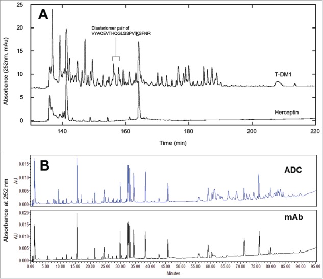
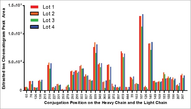
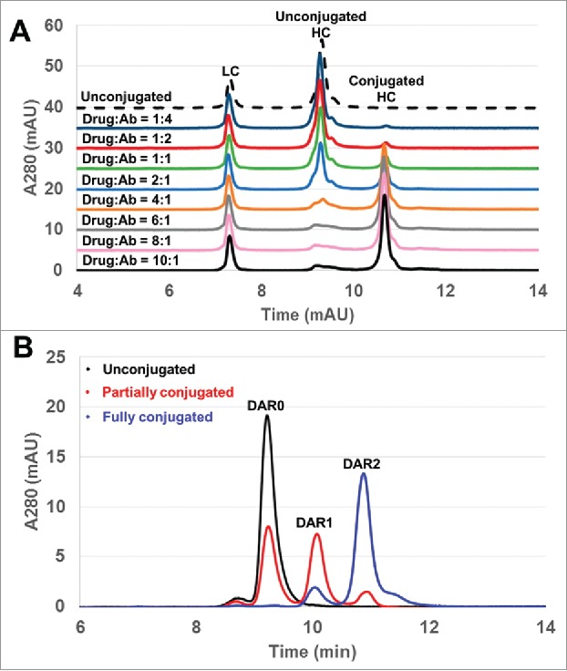
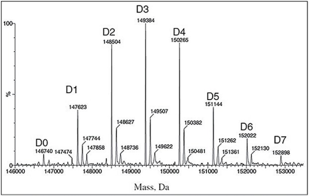
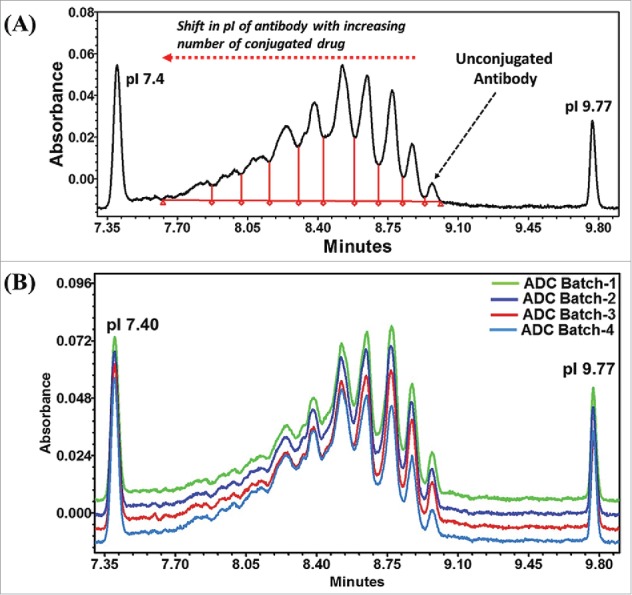
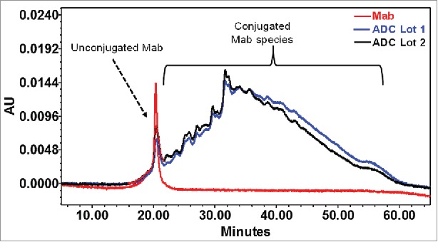
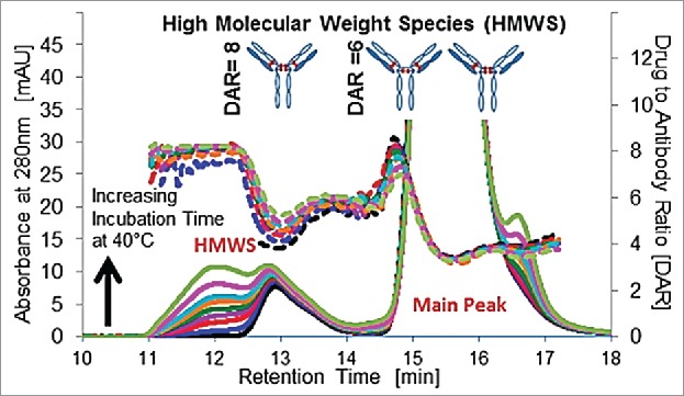
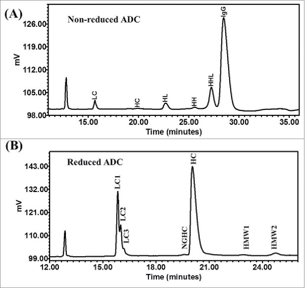
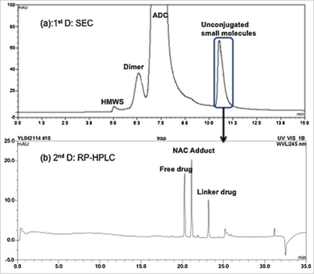
Antibody-drug conjugates (ADCs) are a growing class of biotherapeutics in which a potent small molecule is linked to an antibody. ADCs are highly complex and structurally heterogeneous, typically containing numerous product-related species. One of the most impactful steps in ADC development is the identification of critical quality attributes to determine product characteristics that may affect safety and efficacy. However, due to the additional complexity of ADCs relative to the parent antibodies, establishing a solid understanding of the major quality attributes and determining their criticality are a major undertaking in ADC development. Here, we review the development challenges, especially for reliable detection of quality attributes, citing literature and new data from our laboratories, highlight recent improvements in major analytical techniques for ADC characterization and control, and discuss newer techniques, such as two-dimensional liquid chromatography, that have potential to be included in analytical control strategies.

Keywords: Antibody-drug conjugates; CQA, DAR; analytical; characterization; quality attribute; stability.


Figures

Figure 1.
Figure 1.
Schematic representation of ADCs with different conjugation chemistries.
Figure 2.
Figure 2.
UV-Visible absorbance spectra of (A) free emtansine (DM-1), (B) Trastuzumab, and (C) Trastuzumab emtansine conjugate. The molar concentrations of antibody and drug are determined based on measured absorbance and extinction coefficients (Reprinted with permission, from reference 37).
Figure 3.
Figure 3.
Graphical representation of IdeS cleavage, 2-MEA reduction, and cathepsin B cleavage of ADC. Reprinted with permission, from reference 55.
Figure 4.
Figure 4.
(A) Deconvoluted mass spectrum for the deglycosylated mcMMAF conjugate, ADC-A, and the corresponding parent material, Mab-A (reference 67) (B) Deconvoluted mass spectrum of a deglycosylated mc-vc-tubulysine conjugate.
Figure 5.
Figure 5.
Hydrophobic interaction chromatography (HIC) of Glyco ADCs prepared with 3 different sialylated antibodies and 2 different aminooxy drug-linkers (AO-MMAE and AO-PEG8 Dol10). (A) Trastuzumab conjugated with AO-MMAE. (B) Trastuzumab conjugated with AO-PEG8-Dol10. (C) Anti-fibroblast activation protein (FAP) antibody B11 conjugated with AO-MMAE. (D) Anti-FAP antibody G11 conjugated with AO-MMAE. (E) Trastuzumab conjugated with MC-VC-PABC-MMAE (thiol conjugate). Numbers below each peak indicate the number of drugs conjugated. AO-MMAE: Aminooxy-Cys-MC-VC-PABC-MMAE; AO-PEG8-Dol10: Aminooxy-Cys-MC-VCPABC-PEG8-Dol10. (Reprinted with permission, from reference 70).
Figure 6.
Figure 6.
(A) Overlaid UV traces at 252 nm of trastuzumab emtansine and Herceptin sample tryptic digests separated by reversed-phase HPLC. An example pair of diasteriomer peaks, corresponding to the same conjugation site, is annotated. Only the portions of each chromatogram showing the elution region of the drug-containing peptides are presented in the figure. (reference 46). (B) Overlaid UV traces at 252 nm of a lysine mc-vc-tubulysine conjugate and the correspondent antibody tryptic digests separated by reversed-phase UPLC.
Figure 7.
Figure 7.
Peak areas in the extracted ion chromatograms of all detected conjugated peptides in tryptic digest of a lysine mc-vc-tubulysine random conjugate. Four lots produced by the same process were analyzed side-by-side to demonstrate process consistency.
Figure 8.
Figure 8.
(A) DAR for complete conjugation. Using the reduced RP-HPLC method as a monitoring tool, a range of drug−antibody molar ratios from 1:4 to 10:1 were used for MMAF conjugation on HC-F404. The conjugations were performed at room temperature overnight. (B) Intact DAR characterization by RP-HPLC. RP-HPLC chromatograms revealed baseline resolution of DAR0, DAR1, and DAR2 species for unconjugated (black), partially conjugated (red) and fully conjugated (blue) HC-F404-MMAF. (Reprinted with permission, from reference 74).
Figure 9.
Figure 9.
Mass spectrum of deglycosylated huc242-DM4 conjugate. The peak labels indicate the number of DM4 molecules attached to the antibody: D0 is the unconjugated antibody, while Dn corresponds to antibody molecules carrying n DM4 molecules (for n = 1, 2,…, 8) (Reprinted with permission, from reference 41).
Figure 10.
Figure 10.
(A) Electropherogram of a model Lysine based ADC. The amount of unconjugated antibody was determined by using calibration curve. (B) ICIEF as a finger printing technique to monitor the batch-to-batch process consistency.
Figure 11.
Figure 11.
HIC profile of lysine-conjugated ADCs lot 1 (black) and lot 2 (blue), yielding one prominent peak corresponding to unconjugated antibody (red) and several unresolved peaks for drug-conjugated antibody species. The estimated level of unconjugated antibody was around 4%.
Figure 12.
Figure 12.
SEC chromatograms (at 280 nm) of T = 0 through 8 weeks at 40°C DAR 3.5 ADC stability samples with DAR shown (dotted lines) on the right axis for the HMWS and main peak. Reprinted with permission, from reference 85.
Figure 13.
Figure 13.
Analysis of lysine-conjugated ADCs by CE-SDS under non-reduced (A) and reduced (B) conditions. Peak identification: Internal standard (10 kDa), Light chain (LC), Non-glycosylated heavy chain (NGHC), Heavy chain (HC), 2 Heavy chains (HH), 2 heavy 1 light chain (HHL), IgG monomer.
Figure 14.
Figure 14.
Analysis of unconjugated small molecules in ADC by 2D-LC. (a) SEC on 1st D that separates the unconjugated small molecules from the ADC, and (b) RP-HPLC on 2nd D that separates the three unconjugated small molecules. Reprinted with permission, from reference 84.

Similar articles

Cited by

References

    1. Coulson A, Levy A, Gossell-Williams M. Monoclonal Antibodies in Cancer Therapy: Mechanisms, Successes and Limitations. West Indian Med J. 2014;63(6):650–4. doi:10.7727/wimj.2013.241. PMID:25803383. - DOI - PMC - PubMed
    1. Reichert JM. Antibodies to watch in 2017. MAbs. 2017;9:167–81. doi:10.1080/19420862.2016.1269580. PMID:27960628. - DOI - PMC - PubMed
    1. Leyens L, Brand A. Early Patient Access to Medicines: Health Technology Assessment Bodies Need to Catch Up with New Marketing Authorization Methods. Public Health Genomics. 2016;19:187–91. doi:10.1159/000446537. PMID:27238553. - DOI - PMC - PubMed
    1. Parslow AC, Parakh S, Lee FT, Gan HK, Scott AM. Antibody-Drug Conjugates for Cancer Therapy. Biomedicines. 2016;4(3). pii: E14. doi:10.3390/biomedicines4030014. PMID:28536381. - DOI - PMC - PubMed
    1. Tolcher AW. Antibody drug conjugates: lessons from 20 years of clinical experience. Ann Oncol. 2016;27:2168–72. doi:10.1093/annonc/mdw424. PMID:27733376. - DOI - PubMed

Substances