. 2018 Jan 2;13(1):e0190307.
doi: 10.1371/journal.pone.0190307. eCollection 2018.

Pharmacological activation of TRPV4 produces immediate cell damage and induction of apoptosis in human melanoma cells and HaCaT keratinocytes

Affiliations

Pharmacological activation of TRPV4 produces immediate cell damage and induction of apoptosis in human melanoma cells and HaCaT keratinocytes

Aida Olivan-Viguera et al. PLoS One. .

Abstract

Background: TRPV4 channels are calcium-permeable cation channels that are activated by several physicochemical stimuli. Accordingly, TRPV4 channels have been implicated in the regulation of osmosensing, mechanotransduction, thermosensation, and epithelial/endothelial barrier functions. Whether TRPV4 is also mechanistically implicated in melanoma cell proliferation is not clear. Here, we hypothesized that TRPV4 is expressed in human melanoma and that pharmacological activation interferes with cell proliferation.

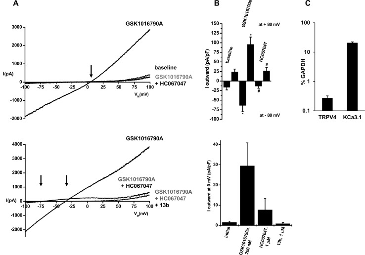
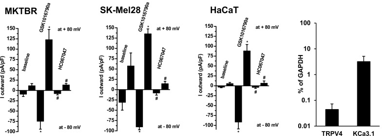
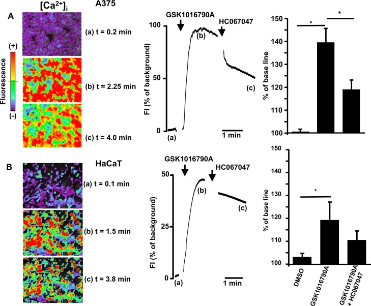
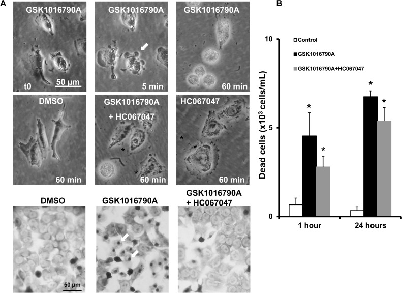
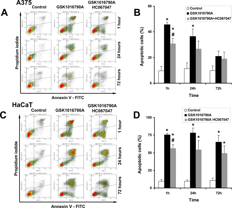
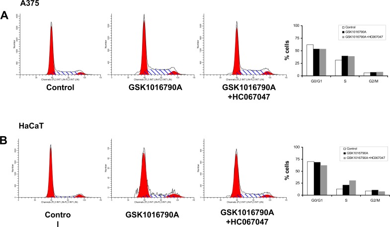
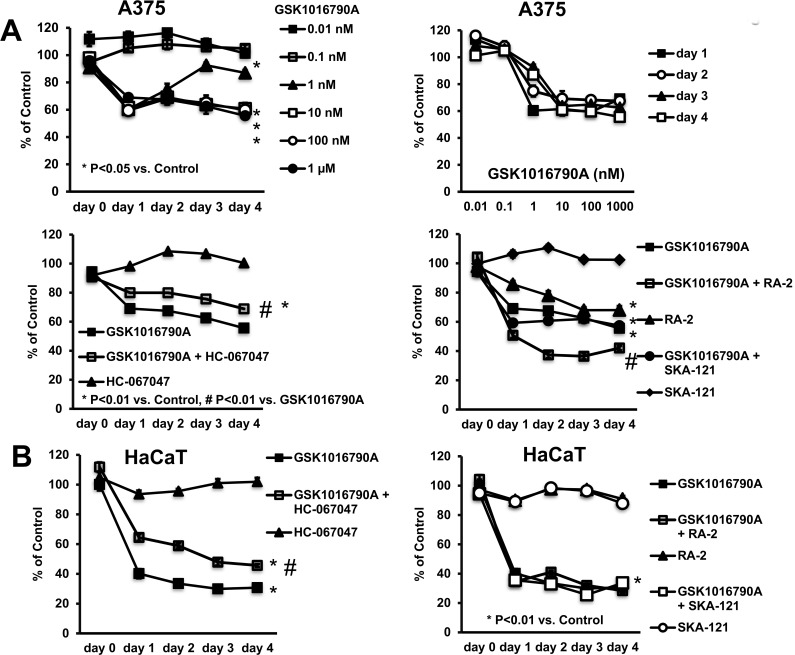
Methodology/principal findings: TRPV4 functions were studied in melanoma cell lines (A375, SK-MEL-28, MKTBR), immortalized non-cancer keratinocytes (HaCaT), and murine 3T3 fibroblasts by patch-clamp, qRT-PCR, intracellular calcium measurements, cell proliferation, and flow cytometric assays of apoptosis and cell cycle. The selective TRPV4-activator, GSK1016790A, elicited non-selective cation currents with TRPV4-typical current-voltage-relationship in all cell lines. GSK1016790A-induced currents were blocked by the TRPV4-blocker, HC067047. TRPV4 mRNA expression was demonstrated by qRT-PCR. In A375 cells, TRPV4 activation was frequently paralleled by co-activation of calcium/calmodulin-regulated KCa3.1 channels. Light microscopy showed that TRPV4-activation produced rapid cellular disarrangement, nuclear densification, and detachment of a large fraction of all melanoma cell lines and HaCaT cells. TRPV4-activation induced apoptosis and drastically inhibited A375 and HaCaT proliferation that could be partially prevented by HC067047.

Conclusions/significance: Our study showed that TRPV4 channels were functionally expressed in human melanoma cell lines and in human keratinocytes. Pharmacological TRPV4 activation in human melanoma cells and keratinocytes caused severe cellular disarrangement, necrosis and apoptosis. Pharmacological targeting of TRPV4 could be an alternative or adjuvant therapeutic strategy to treat melanoma progression and other proliferative skin disorders.


Conflict of interest statement

Competing Interests: The authors have declared that no competing interests exist.

Figures

Fig 1
Fig 1. Characterization of TRPV4 channels in A375 melanoma cells.
A) Upper panel: Exemplary whole-cell recordings showing activation of TRPV4 channels by GSK1016790A (200 nM) and inhibition of currents by HC067047 (1 μM). The arrow indicates a positive reversal potential of GSK1016790A-activated currents. Baseline currents were not considerable inhibited by HC067047. Lower panel: Co-activation of KCa-currents. The right arrow indicates a negative reversal potential (Erev) of ca. -35 mV of the mixed TRPV4 and KCa current and the left arrow indicates an Erev of ca. -75 mV of the isolated KCa-current after inhibition of TRPV4 currents by HC067047. The KCa-current was fully blocked by the negative-gating modulator of KCa3.1 channels, 13b (1 μM). B) Upper panel: Mean normalized currents at clamp potentials of -80 and +80 mV before and after addition GSK1016790A (n = 8, experiments) and after addition of HC067047 (n = 8). Lower panel: Mean mixed TRPV4/KCa3.1 currents at a clamp potential of 0 mV after addition of GSK1016790A (n = 5) and inhibition of TRPV4 currents by HC067047 (n = 4) and of KCa3.1 currents by 13b (n = 5). C) Quantitative RT-PCR analysis of TRPV4 and KCa3.1 gene expression as percentage of GAPDH expression (replicates, n = 3). Data points are means ± SEM.
Fig 2
Fig 2. GSK1016790A-induced TRPV4-currents and inhibition by HC067047 in the melanoma lines, MKTBR and SK-MEL-28, and the human non-cancer keratinocyte line, HaCaT.
Data points are means ± SEM (cells, n = 4–6 each). Panel on right: Quantitative RT-PCR analysis of TRPV4 and KCa3.1 gene expression in HaCaT as percentage of GAPDH expression (replicates, n = 3). Data points are means ± SEM.
Fig 3
Fig 3
A) Changes of intracellular calcium ([Ca2+i]) in response to TRPV4 activation in A375 cells. Exemplary images of GSK1016790A(1 μM)-induced increase of [Ca2+i] (upper left panel), its time course (upper panel in middle), and (on right) summary data of % change of fluorescence to GSK1016790A (n = 10 independent experiments) and in combination with HC067047 (n = 5 independent experiments). HC067047 (10 μM) partially reversed the response. Note that artifacts caused by compound addition were removed. No change of fluorescence was recorded in the presence of vehicle (DMSO 0.1%, n = 3). B) Lower panel on left: Exemplary images of changes of intracellular calcium ([Ca2+]i) to TRPV4 activation in HaCaT cells. Note that artefacts caused by compound addition to the bath were removed. In middle: Time course of changes in [Ca2+]i in response to GSK1016790A (n = 4 independent experiments) alone and in combination with HC067047 (n = 4 independent experiments). The vehicle (DMSO 0.1%, n = 3) produced virtually no response. On right: Summary of data. Data are means ± SEM; *P<0.001, Student’s T test.
Fig 4
Fig 4. Alterations of cell morphology, cell detachment and cell death induced by GSK1016790A.
A) Upper two panels: Exemplary light microscopic images illustrating time course of cell retraction, membrane blebbing (indicated by arrows), and cell detachment during the first hour of exposure to GSK1016790A (1 μM). HC067047 (1 μM) prevented visibly GSK1016790A-induced changes. HC067047 or vehicle (DMSO) had no visible effect. Lower panel: Giemsa-stained A375 cells after 1 h exposure to GSK1016790A, in combination with HC067047, or DMSO. Note the densification of nuclei (dark-grey dots indicated by arrows) in GSK1016790A-treated cells. B) Counts of non-viable, “death” cells in supernatant. Data points are means ± SEM (number of independent experiments, n = 3). *P<0.05 vs. DMSO, #P <0.05 vs. GSK1016790A; Student’s T test.
Fig 5
Fig 5. FACS analysis of apoptosis.
A) Representative flow cytometry dot plots with double Annexin V-FITC/PI staining for control cells (DMSO 0,2%), cells exposed to GSK1016790A (10 nM), and cells exposed to GSK1016790A and HC067047 (1 μM) at 1 h, 24 h, and 72 h. B) Summary data. C) Induction of apoptosis in HaCaT cells and protective effects of HC067047. D) Summary data. *P<0.05 vs. Control, #P<0.05 vs. GSK1016790A, ANOVA, n = 3). Data are means ± SEM (number of independent experiments, n = 3).
Fig 6
Fig 6
Pharmacological TRPV4 activation did not impair the cell cycle in A375 cells (A) and HaCaT keratinocytes (B).
Fig 7
Fig 7. Impact of GSK1016790A on cell proliferation/survival.
A) Upper panel on left: Concentration-dependent reduction of cell proliferation/survival of A375 as measured by Janus Green-Assay. Upper panel on right: Note that half-maximal inhibition was achieved at ca. 1 nM GSK1016790A for all time intervals, except day-1. Lower panel on left: HC067047 antagonized the response to GSK1016790A. Lower panel on right: The negative gating-modulator of KCa3.1, the 13b derivate, RA-2 (10 μM) reduced cell proliferation/survival and potentiated the response to GSK1016790A. The positive-gating modulator of KCa3.1, SKA-121, had no effects. Data points are means ± SEM (n = 6–36 from n = 2–6 independent experiments). B) GSK1016790A impaired proliferation/survival of HaCaT cells. HC067047 partially antagonized the response. The negative and positive KCa3.1-gating modulators, RA-2 and SKA-121, respectively, did not modulate the response. Data points are means ± SEM (n = 18; number of independent experiments, n = 3). *P<0.05 vs. DMSO, #P<0.05 vs. GSK1016790A; Student’s T test.

References

    1. Carvacho I (2016) Transient Receptor Potential channels: TRPV4. In: Clapham DE, editor: IUPHAR/BPS Guide to PHARMACOLOGY.
    1. White JP, Cibelli M, Urban L, Nilius B, McGeown JG, Nagy I (2016) TRPV4: Molecular Conductor of a Diverse Orchestra. Physiol Rev 96: 911–973 doi: 10.1152/physrev.00016.2015 - DOI - PubMed
    1. Liedtke W, Friedman JM (2003) Abnormal osmotic regulation in trpv4-/- mice. Proc Natl Acad Sci U S A 100: 13698–13703 doi: 10.1073/pnas.1735416100 - DOI - PMC - PubMed
    1. Strotmann R, Harteneck C, Nunnenmacher K, Schultz G, Plant TD (2000) OTRPC4, a nonselective cation channel that confers sensitivity to extracellular osmolarity. Nat Cell Biol 2: 695–702 doi: 10.1038/35036318 - DOI - PubMed
    1. Guler AD, Lee H, Iida T, Shimizu I, Tominaga M, Caterina M (2002) Heat-evoked activation of the ion channel, TRPV4. J Neurosci 22: 6408–6414 - PMC - PubMed

Publication types

Substances