. 2018 Feb 1;141(2):472-485.
doi: 10.1093/brain/awx338.

A widespread visually-sensitive functional network relates to symptoms in essential tremor

Affiliations

A widespread visually-sensitive functional network relates to symptoms in essential tremor

Derek B Archer et al. Brain. .

Abstract

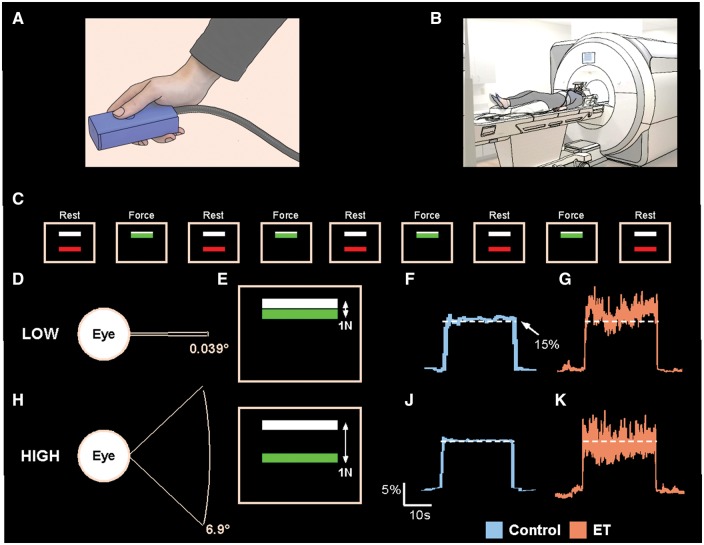
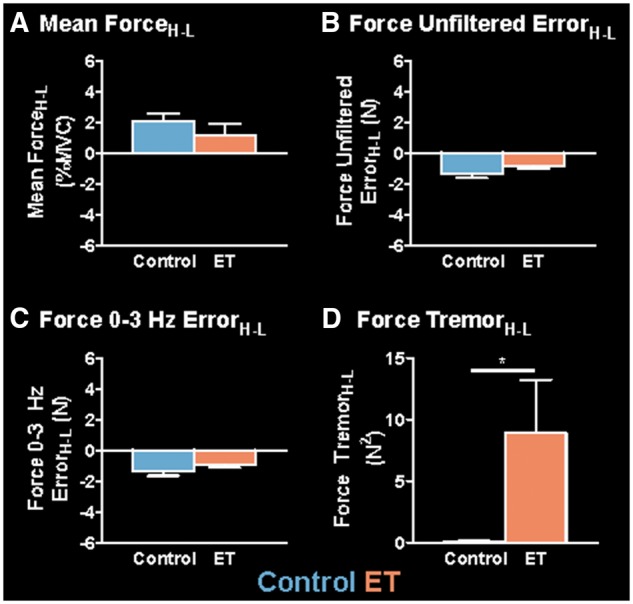
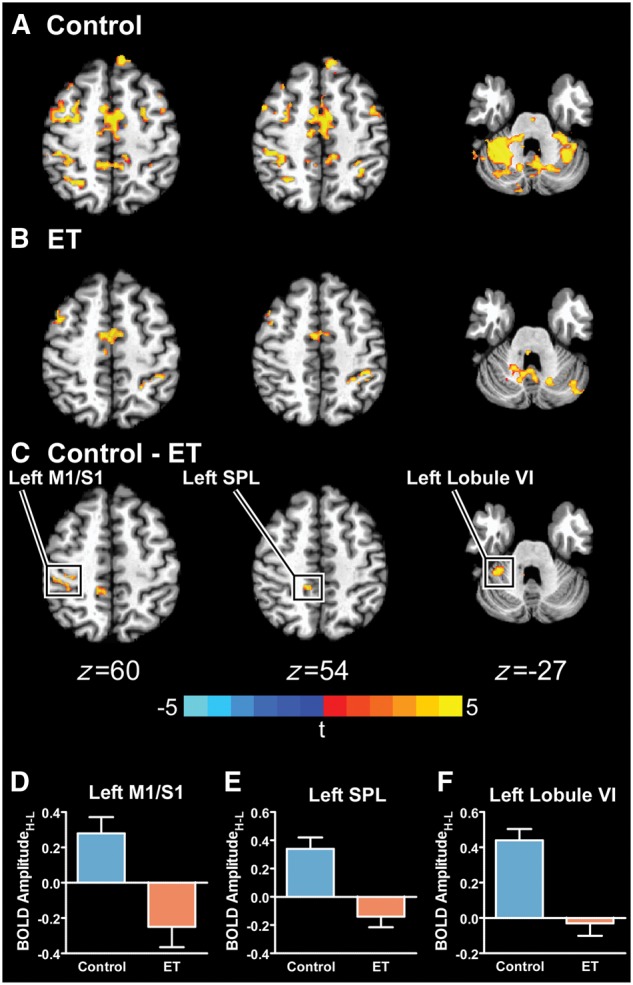
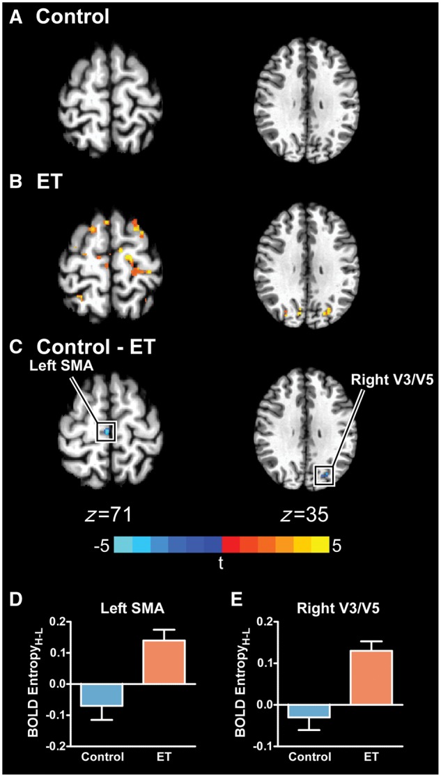
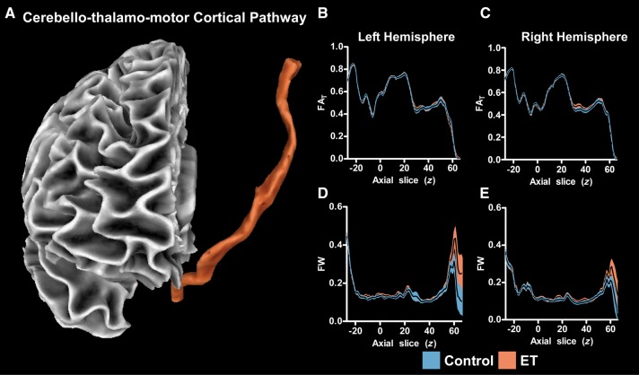
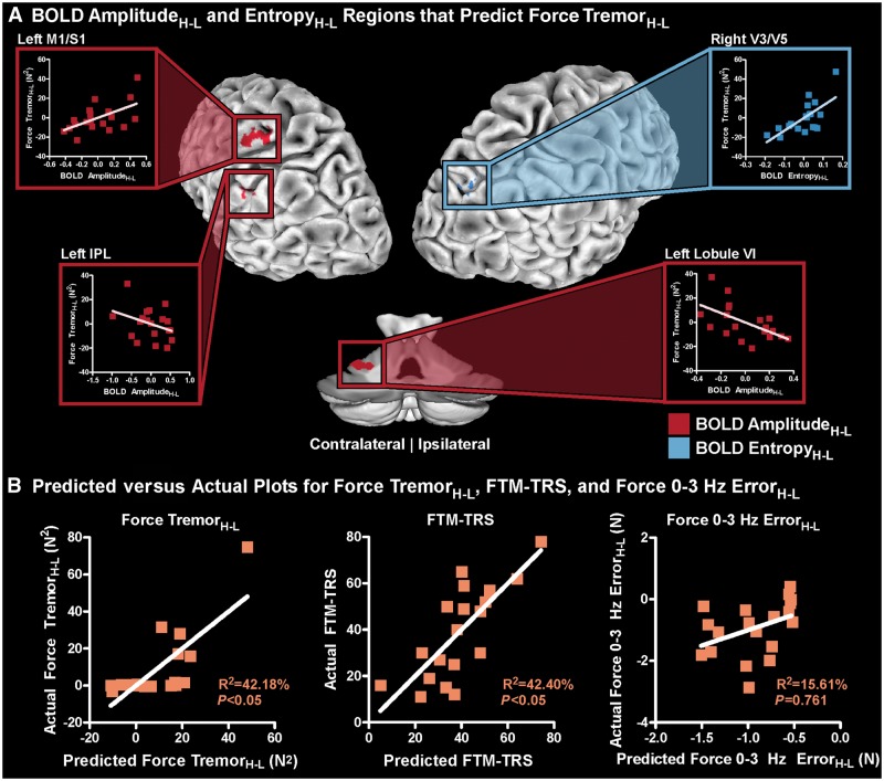
Essential tremor is a neurological syndrome of heterogeneous pathology and aetiology that is characterized by tremor primarily in the upper extremities. This tremor is commonly hypothesized to be driven by a single or multiple neural oscillator(s) within the cerebello-thalamo-cortical pathway. Several studies have found an association of blood-oxygen level-dependent (BOLD) signal in the cerebello-thalamo-cortical pathway with essential tremor, but there is behavioural evidence that also points to the possibility that the severity of tremor could be influenced by visual feedback. Here, we directly manipulated visual feedback during a functional MRI grip force task in patients with essential tremor and control participants, and hypothesized that an increase in visual feedback would exacerbate tremor in the 4-12 Hz range in essential tremor patients. Further, we hypothesized that this exacerbation of tremor would be associated with dysfunctional changes in BOLD signal and entropy within, and beyond, the cerebello-thalamo-cortical pathway. We found that increases in visual feedback increased tremor in the 4-12 Hz range in essential tremor patients, and this increase in tremor was associated with abnormal changes in BOLD amplitude and entropy in regions within the cerebello-thalamo-motor cortical pathway, and extended to visual and parietal areas. To determine if the tremor severity was associated with single or multiple brain region(s), we conducted a birectional stepwise multiple regression analysis, and found that a widespread functional network extending beyond the cerebello-thalamo-motor cortical pathway was associated with changes in tremor severity measured during the imaging protocol. Further, this same network was associated with clinical tremor severity measured with the Fahn, Tolosa, Marin Tremor Rating Scale, suggesting this network is clinically relevant. Since increased visual feedback also reduced force error, this network was evaluated in relation to force error but the model was not significant, indicating it is associated with force tremor but not force error. This study therefore provides new evidence that a widespread functional network is associated with the severity of tremor in patients with essential tremor measured simultaneously at the hand during functional imaging, and is also associated with the clinical severity of tremor. These findings support the idea that the severity of tremor is exacerbated by increased visual feedback, suggesting that designers of new computing technologies should consider using lower visual feedback levels to reduce tremor in essential tremor.

Keywords: cerebellar function; motor control; motor cortex; movement disorders; tremor.


Figures

Figure 1
Figure 1
Experimental set-up. The force transducer was held between the thumb and the index finger by the participant during the MRI session (A), and the participant laid in the supine position in which the hand and transducer rested on the upper leg (B). Above the field of view of the participant was a mirror, which reflected the visual display. The visual display instructed the participant when to produce force. Initially, the participant rested for 30 s in the ‘Rest’ condition (C). At the end of the Rest condition, the colour of the bar would change, indicating the beginning of the ‘Force’ condition. The participant then began producing force for 30 s, thus fluctuating around the white target bar. At the end of the 30 s, the colour of the bar would change again, indicating the beginning of the ‘Rest’ condition. An example showing the visual gain manipulation (D and H), how it is perceived by the subject on the visual display (E and I), and example force traces for a control subject (F and J) and an essential tremor (ET) subject (G and K) is also shown for the low and high visual feedback conditions.
Figure 2
Figure 2
Group differences in mean ForceH-L, Force Unfiltered ErrorH-L, Force 0–3 Hz ErrorH-L, and Force TremorH-L. Mean values for each measure are shown for the controls (blue) and essential tremor (ET) patients (orange). Mean ForceH-L (A), Force Unfiltered ErrorH-L (B), Force 0–3 Hz ErrorH-L (C), and Force TremorH-L (D) are shown for the controls (blue) and essential tremor patients (orange). Each bar represents the group mean at each level of visual feedback, and error bars represent ±standard error of the mean (SEM). *Significant between-group differences (P < 0.05).
Figure 3
Figure 3
Between-group BOLD AmplitudeH-L differences. The significant BOLD AmplitudeH-L for controls (A), essential tremor (ET) patients (B), and the between-group difference (PFWER < 0.005) (C) is shown. The boxed regions in C are plotted in D–F, in which the mean BOLD AmplitudeH-L for controls (blue) and essential tremor patients (orange) is shown. Errors bars represent ± SEM.
Figure 4
Figure 4
Between-group BOLD EntropyH-L differences. The significant BOLD EntropyH-L for controls (A), essential tremor (ET) patients (B), and the between-group difference (PFWER < 0.005) (C) is shown. The boxed regions in C are plotted in D and E, in which the mean BOLD EntropyH-L for controls (blue) and essential tremor patients (orange) is shown. Errors bars represent ± SEM. SMA = supplementary motor area.
Figure 5
Figure 5
Probabilistic tractography FAT and free water profiles. A 3D view of the cerebello-thalamo-motor cortical tract is shown (A). The FAT profiles are shown for the left (B) and right (C) hemispheres. The mean FAT is displayed with a black line, and the blue (control) and orange (essential tremor, ET) shaded areas represent ± SEM for each group. The free water (FW) profiles are also shown for the left (D) and right (E) hemispheres. No significant between-group differences in FAT or free water were found.
Figure 6
Figure 6
Multiple regression to associate Force TremorH-L with BOLD signal. (A) The BOLD AmplitudeH-L and EntropyH-L regions, which were associated with Force TremorH-L includes left M1/S1, IPL, cerebellar lobule VI, and right V3/V5. BOLD AmplitudeH-L and EntropyH-L regions are shown in red and blue, respectively. For each region, a partial regression plot is displayed, which shows how each region contributes to the multiple regression model while controlling for all other variables. (B) The predicted versus actual plots for Force TremorH-L, FTM-TRS, and Force 0-3 Hz ErrorH-L. Age was included as a behavioural covariate.

Comment in

References

    1. Andersson JLR, Sotiropoulos SN. An integrated approach to correction for off-resonance effects and subject movement in diffusion MR imaging. Neuroimage 2016; 125: 1063–78. - PMC - PubMed
    1. Archer DB, Misra G, Patten C, Coombes SA. Microstructural properties of premotor pathways predict visuomotor performance in chronic stroke. Hum Brain Mapp 2016; 37: 2039–54. - PMC - PubMed
    1. Archer DB, Vaillancourt DE, Coombes SA. A template and probabilistic atlas of the human sensorimotor tracts using diffusion MRI. Cereb Cortex 2017; 1–15. doi: 10.1093/cercor/bhx066. - PMC - PubMed
    1. Avants BB, Epstein CL, Grossman M, Gee JC. Symmetric diffeomorphic image registration with cross-correlation: evaluating automated labeling of elderly and neurodegenerative brain. Med Image Anal 2008; 12: 26–41. - PMC - PubMed
    1. Barbe MT, Franklin J, Kraus D, Reker P, Dembek TA, Allert N, et al.Deep brain stimulation of the posterior subthalamic area and the thalamus in patients with essential tremor: study protocol for a randomized controlled pilot trial. Trials 2016; 17: 476. - PMC - PubMed

Publication types

MeSH terms