. 2018 Mar 16;46(5):2585-2599.
doi: 10.1093/nar/gkx1286.

Translational regulation by bacterial small RNAs via an unusual Hfq-dependent mechanism

Affiliations

Translational regulation by bacterial small RNAs via an unusual Hfq-dependent mechanism

Muhammad S Azam et al. Nucleic Acids Res. .

Abstract

In bacteria, the canonical mechanism of translational repression by small RNAs (sRNAs) involves sRNA-mRNA base pairing that occludes the ribosome binding site (RBS), directly preventing translation. In this mechanism, the sRNA is the direct regulator, while the RNA chaperone Hfq plays a supporting role by stabilizing the sRNA. There are a few examples where the sRNA does not directly interfere with ribosome binding, yet translation of the target mRNA is still inhibited. Mechanistically, this non-canonical regulation by sRNAs is poorly understood. Our previous work demonstrated repression of the mannose transporter manX mRNA by the sRNA SgrS, but the regulatory mechanism was unknown. Here, we report that manX translation is controlled by a molecular role-reversal mechanism where Hfq, not the sRNA, is the direct repressor. Hfq binding adjacent to the manX RBS is required for sRNA-mediated translational repression. Translation of manX is also regulated by another sRNA, DicF, via the same non-canonical Hfq-dependent mechanism. Our results suggest that the sRNAs recruit Hfq to its binding site or stabilize the mRNA-Hfq complex. This work adds to the growing number of examples of diverse mechanisms of translational regulation by sRNAs in bacteria.


Figures

Figure 1.
Figure 1.
SgrS-mediated translational repression of manX is Hfq-dependent. (A) Strain JH175 has a manX′-′lacZ translational fusion under the control of Cp19, a constitutive promoter. This strain was transformed with a vector control or plasmids with SgrS homologs from Salmonella (SgrSSal) or E. coli (SgrSEco), induced with 0.1 mM IPTG, and assayed for β-galactosidase activity after 60 min. Units of activity in the experimental samples were normalized to the levels in the vector control strains to yield percent relative activity. (B) The same vector control and SgrSSal plasmids described in A were transformed to strain JH184 (hfq+) or SA1705 (Δhfq), containing a Cp19-ptsG′-′lacZ fusion. Induction of SgrS and β-Galactosidase assays were conducted and analyzed as in A. (C) Strains JH175 (hfq+) and SA1328 (Δhfq) with the Cp19- manX′-′lacZ fusion were transformed with vector control or SgrSSal plasmids. SgrS was induced using the indicated concentrations of IPTG and β-galactosidase assays were conducted and analyzed as in A.
Figure 2.
Figure 2.
Genetic analysis of a putative Hfq binding site in the manX 5′ UTR. (A) The full-length manX UTR in E. coli is 115-nt. Four translation fusions with truncations of the UTR (as indicated) were constructed by moving the heterologous promoter closer to the TIR. Vector control and SgrS plasmids were transformed into the resulting strains (JH178, JH181, SA1404 and SA1403) and β-Galactosidase assays were conducted and analyzed as described for Figure 1A. (B) An A/U rich motif upstream of the manX RBS, was mutated in the context of the 25-nt manX translational fusion, resulting in the mut-1 fusion. The positions of the putative Hfq binding site and confirmed SgrS binding site are indicated with gray boxes. The RBS is in blue letters and the manX start codon is in green. The activity of SgrS on wild-type (strain SA1404) and mut-1 (strain SA1522) fusions was assessed after induction as described for Figure 1A. (C) Additional mutations as indicated in red were constructed in the putative Hfq binding site in the manX translational fusion, resulting in mutant fusions mut-2 through mut-5 (strains SA1713, SA1711, SA1712, SA1710, respectively). The plasmids, induction and β-Galactosidase assays were conducted and analyzed as described for Figure 1A, except that activities are reported in Miller Units.
Figure 3.
Figure 3.
Mutations Hfq impact translation and stability of manX mRNA. (A) Wild-type and mut-1 through mut-5manX translational fusions (same as Figure 2C) in hfq+ (+) and Δhfq (Δ) backgrounds (for strains, see Supplementary Table S1) were assayed for β-galactosidase activity. Cultures were grown to mid-log phase and then assayed. Specific activity is reported in Miller Units. (B) The rne131 allele was moved into the reporter strains described in A. β-Galactosidase activity was assayed as described in A.
Figure 4.
Figure 4.
Toeprint assays reveal that Hfq, but not SgrS, can prevent ribosome binding to manX mRNA. (A) Toeprint assays were conducted as described in Materials and Methods. Ribosomes, tRNAfMet and Hfq were added to manX mRNA as indicated above the gel image. In lanes 4–6, Hfq concentrations were 0.15, 0.5 and 1 μM, respectively. The sequencing ladder is indicated by ‘T – G – C – A’, and was generated with the same oligo (OJH119) used for reverse transcription. The toeprint signal is indicated at +15/16 relative to the start codon. (B) Lanes 1–6 on the left represent the same reactions as described in part A. Lanes 7–12 on the right represent similar reactions, except SgrS was added at concentrations of 100, 250 and 500 nM (lanes 10–12).
Figure 5.
Figure 5.
DicF, a prophage-encoded sRNA, also regulates manX translation. (A) Strains with manX′-′lacZ fusions with a wild-type (JH175) or mutant (putative) Hfq binding site mut-6 (SA1620, with the same Hfq binding site mutation as mut-1, but the fusion contains more manX coding sequence to include the DicF binding site) (as shown in Figure 2B) were transformed with vector control or DicF-expressing plasmids. Expression of DicF was induced with 0.1 mM IPTG, and β-Galactosidase assays were conducted and analyzed as described for Figure 1A. (B) Base pairing interactions for manX mRNA (middle sequence) and SgrS (top sequence) or DicF (bottom sequence). The position of the DicF20 mutation is indicated below the DicF sequence. (C) Strain JH175, containing the wild-type manX′-′lacZ fusion, was transformed with vector control or plasmids expressing wild-type DicF or mutant DicF20. Expression of DicF was induced with 0.1 mM IPTG, and β-galactosidase assays were conducted and analyzed as described for Figure 1A.
Figure 6.
Figure 6.
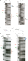
Footprinting maps SgrS, DicF and Hfq binding sites on manX RNA. In vitro transcribed manX mRNA containing the full-length 115-nt UTR and a portion of the coding region extending 51 nt downstream of the predicted DicF base pairing region was end labeled with 32P and incubated with and without unlabeled SgrS, DicF and Hfq to perform footprinting reactions. Samples were treated as follows: ‘T1,’ RNase T1; ‘OH,’ alkaline ladder; ‘PbAc,’ lead acetate. Positions of G residues are indicated to the left of each gel image and nucleotides numbered as indicated in B. Positions of the GUG start codon and RBS are indicated to the left of each image. (A) Footprinting SgrS and Hfq (left image) or DicF and Hfq (right image) binding sites on manX mRNA. (B) Sequence and putative structure of manX following interaction with Hfq and SgrS or DicF. Positions of SgrS and DicF binding are indicated (from residues 139–167). The start codon is indicated by orange nucleotides. The Hfq binding site is highlighted in orange and the RBS is highlighted in green. (C and D) Footprinting using reduced concentrations of Hfq (as described in Materials and Methods) in the absence and presence of SgrS. manX with wild-type Hfq binding motif, AUAAUAAA is shown in C and the mut-1 site, CGGCGGGA, is shown in D.
Figure 7.
Figure 7.
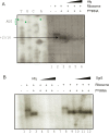
In vitro analyses of sRNA binding to manX mRNA and Hfq. (A) Native gel electrophoresis was used to examine binding of manX mRNA with SgrS and DicF sRNAs. In vitro transcribed 32P-labeled manX mRNA (0.01 pmol) was mixed with indicated amounts of cold SgrS and incubated at 37°C for 30 min. The reaction mixture was resolved on a chilled native acrylamide gel. Bands were quantified and the fraction of manX mRNA bound was calculated and plotted to calculate KD. (B) Gel mobility shift assay for SgrS (right) or DicF (left) and Hfq. Measured band densities (n replicates, top left) were plotted to determine the dissociation constants.
Figure 8.
Figure 8.
A model for the non-canonical roles played by two distinct sRNAs, SgrS and DicF, to repress manX translation via an Hfq-dependent mechanism.

References

    1. Tomizawa J., Itoh T., Selzer G., Som T.. Inhibition of ColE1 RNA primer formation by a plasmid-specified small RNA. Proc. Natl. Acad. Sci. U.S.A. 1981; 78:1421–1425. - PMC - PubMed
    1. Stougaard P., Molin S., Nordstrom K.. RNAs involved in copy-number control and incompatibility of plasmid R1. Proc. Natl. Acad. Sci. U.S.A. 1981; 78:6008–6012. - PMC - PubMed
    1. Morfeldt E., Taylor D., von Gabain A., Arvidson S.. Activation of alpha-toxin translation in Staphylococcus aureus by the trans-encoded antisense RNA, RNAIII. EMBO J. 1995; 14:4569–4577. - PMC - PubMed
    1. Lease R.A., Cusick M., Belfort M.. Riboregulation in Escherichia coli: DsrA RNA acts by RNA:RNA interactions at multiple loci. Proc. Natl. Acad. Sci. U.S.A. 1998; 95:12456–12461. - PMC - PubMed
    1. Frohlich K.S., Papenfort K., Fekete A., Vogel J.. A small RNA activates CFA synthase by isoform-specific mRNA stabilization. EMBO J. 2013; 32:2963–2979. - PMC - PubMed

Publication types

MeSH terms

LinkOut - more resources