

Increased Resting Hippocampal and Basal Ganglia Perfusion in People at Ultra High Risk for Psychosis: Replication in a Second Cohort
- PMID: 29294102
- PMCID: PMC6192497
- DOI: 10.1093/schbul/sbx169
Increased Resting Hippocampal and Basal Ganglia Perfusion in People at Ultra High Risk for Psychosis: Replication in a Second Cohort
Abstract
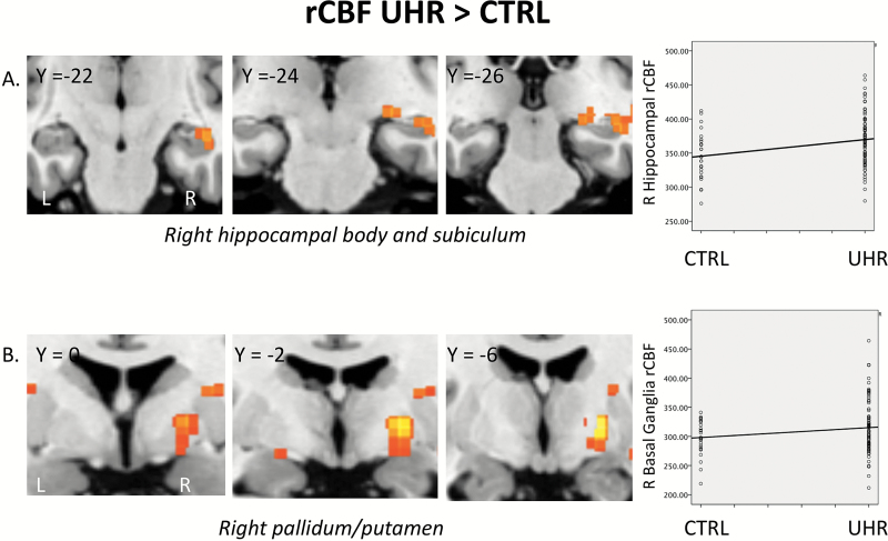
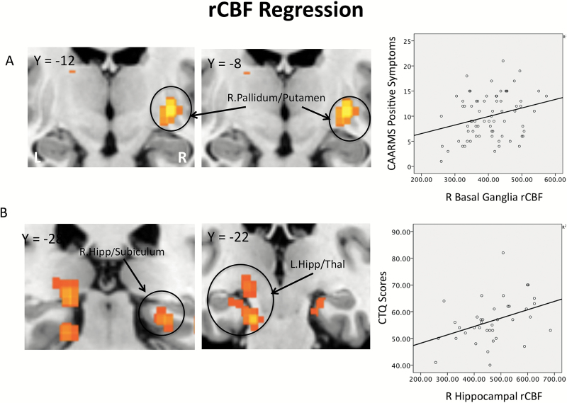
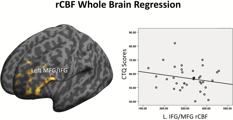
We recently reported that resting hippocampal, basal ganglia and midbrain perfusion is elevated in people at ultra high risk (UHR) for psychosis. The present study sought to replicate our previous finding in an independent UHR cohort, and examined the relationship between resting perfusion in these regions, psychosis and depression symptoms, and traumatic experiences in childhood. Pseudo-Continuous Arterial Spin Labelling (p-CASL) imaging was used to measure resting cerebral blood flow (rCBF) in 77 UHR for psychosis individuals and 25 healthy volunteers in a case-control design. UHR participants were recruited from clinical early detection services at 3 sites in the South of England. Symptoms levels were assessed using the Comprehensive Assessment of At Risk Mental States (CAARMS), the Hamilton Depression Scale (HAM-D), and childhood trauma was assessed retrospectively using the Childhood Trauma Questionnaire (CTQ). Right hippocampal and basal ganglia rCBF were significantly increased in UHR subjects compared to controls, partially replicating our previous finding in an independent cohort. In UHR participants, positive symptoms were positively correlated with rCBF in the right pallidum. CTQ scores were positively correlated with rCBF values in the bilateral hippocampus and negatively associated with rCBF in the left prefrontal cortex. Elevated resting hippocampal and basal ganglia activity appears to be a consistent finding in individuals at high risk for psychosis, consistent with data from preclinical models of the disorder. The association with childhood trauma suggests that its influence on the risk of psychosis may be mediated through an effect on hippocampal function.
Figures
References
-
- Honea R, Crow TJ, Passingham D, Mackay CE. Regional deficits in brain volume in schizophrenia: a meta-analysis of voxel-based morphometry studies. Am J Psychiatry. 2005;162:2233–2245. - PubMed
-
- Tamminga CA, Stan AD, Wagner AD. The hippocampal formation in schizophrenia. Am J Psychiatry. 2010;167:1178–1193. - PubMed
-
- Mechelli A, Riecher-Rössler A, Meisenzahl EM et al. Neuroanatomical abnormalities that predate the onset of psychosis: a multicenter study. Arch Gen Psychiatry. 2011;68:489–495. - PubMed
-
- Pantelis C, Velakoulis D, McGorry PD et al. Neuroanatomical abnormalities before and after onset of psychosis: a cross-sectional and longitudinal MRI comparison. Lancet. 2003;361:281–288. - PubMed
Publication types
MeSH terms
Grants and funding
LinkOut - more resources
Full Text Sources
Other Literature Sources
Medical
