Review
. 2018 Apr;11(2):123-132.
doi: 10.1007/s12265-017-9778-5. Epub 2018 Jan 2.

Atlas-Based Computational Analysis of Heart Shape and Function in Congenital Heart Disease

Affiliations
Review

Atlas-Based Computational Analysis of Heart Shape and Function in Congenital Heart Disease

Kathleen Gilbert et al. J Cardiovasc Transl Res. 2018 Apr.

Abstract

Approximately 1% of all babies are born with some form of congenital heart defect. Many serious forms of CHD can now be surgically corrected after birth, which has led to improved survival into adulthood. However, many patients require serial monitoring to evaluate progression of heart failure and determine timing of interventions. Accurate multidimensional quantification of regional heart shape and function is required for characterizing these patients. A computational atlas of single ventricle and biventricular heart shape and function enables quantification of remodeling in terms of z scores in relation to specific reference populations. Progression of disease can then be monitored effectively by longitudinal evaluation of z scores. A biomechanical analysis of cardiac function in relation to population variation enables investigation of the underlying mechanisms for developing pathology. Here, we summarize recent progress in this field, with examples in single ventricle and biventricular congenital pathologies.

Keywords: Atlas-based analysis; Congenital heart disease; Patient-specific modeling.


Conflict of interest statement

Competing interests

ADM and JHO are a co-founders of, and have an equity interest in Insilicomed, Inc., and serve on the scientific advisory board. ADM is a co-founder of Vektor Medical, Inc., and serves on the scientific advisory board. Some of their research grants, including those acknowledged here, have been identified for conflict of interest management based on the overall scope of the project and its potential benefit to Insilicomed, Inc. The authors are required to disclose this relationship in publications acknowledging the grant support, however the research subject and findings reported here did not involve the company in any way and have no relationship whatsoever to the business activities or scientific interests of the company. The terms of this arrangement have been reviewed and approved by the University of California San Diego in accordance with its conflict of interest policies. The other authors have no competing interests to declare.

Figures

Figure 1
Figure 1
Cardiac magnetic resonance images with epicardial and endocardial contours. a) long axis view b) short axis view and c) 3D biventricular model of a patient with tetralogy of Fallot. d) long axis view e) short axis view and f) 3D single ventricle model.
Figure 2
Figure 2
Four chamber ED frames from three participants showing the three shapes identified. (a) LV dominant, (b) RV dominant (c) double apex. The images are from three participants with, (a) Dextro-Transposition of the great arteries, (b) tetralogy of Fallot, and (c) a ventricular septal defect and coarctation of the aorta.
Figure 3
Figure 3
Pipeline for creating new CHD specific templates and patient specific models. Step 1: 3D CMR data is segmented; Step 2: Notes in the template are defined; Step 3: Subdivision surface give the shape-specific template; Step 4: the template is interactively customized to short and long axis cine images in each frame; Step 5: finite element models are generated for biomechanical analysis.
Figure 4
Figure 4
The shape-specific templates. (a) LV dominant, (b) RV dominant and (c) is double apex. The LV is shown in green, the RV in yellow the biventricular epicardial surface is in blue. MV-mitral valve, AV-aortic valve, TV-tricuspid valve and PV-pulmonary valve.
Figure 5
Figure 5
The first three modes of the ED atlas. The left hand column shows the 5th percentile, the middle column the average shape and the right hand column the 95th percentile. The LV is shown in pink, the RV in green and the biventricular epicardium in blue.
Figure 6
Figure 6
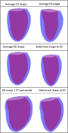
Top: The average shapes at ED and ES for the Fontan dataset. Middle: Average ED shape and the resulting deformed ES shape. Bottom: 5th percentile of mode 1 and the resulting deformed ES shape.

References

    1. Marelli AJ, Mackie AS, Ionescu-Ittu R, Rahme E, Pilote L. Congenital heart disease in the general population: changing prevalence and age distribution. Circulation. 2007;115(2):163–172. doi:CIRCULATIONAHA.106.627224 [pii] - PubMed
    1. Roest AA, de Roos A. Imaging of patients with congenital heart disease. [Review] Nat Rev Cardiol. 2012;9(2):101–115. doi: 10.1038/nrcardio.2011.162. - DOI - PubMed
    1. Geva T. Repaired tetralogy of Fallot: the roles of cardiovascular magnetic resonance in evaluating pathophysiology and for pulmonary valve replacement decision support. J Cardiovasc Magn Reson. 2011;13:9. pii. - PMC - PubMed
    1. McKenzie ED, Khan MS, Dietzman TW, Guzman-Pruneda FA, Samayoa AX, Liou A, et al. Surgical pulmonary valve replacement: a benchmark for outcomes comparisons. J Thorac Cardiovasc Surg. 2014;148(4):1450–1453. doi: 10.1016/j.jtcvs.2014.02.060. - DOI - PubMed
    1. Coats L, O’Connor S, Wren C, O’Sullivan J. The single-ventricle patient population: a current and future concern a population-based study in the North of England. [Research Support, Non-U.S. Gov’t] Heart. 2014;100(17):1348–1353. doi: 10.1136/heartjnl-2013-305336. - DOI - PubMed

Publication types

LinkOut - more resources