. 2017 Dec 23;23(1):32.
doi: 10.3390/molecules23010032.

Structural Basis for the Substrate Inhibition of Proline Utilization A by Proline

Affiliations

Structural Basis for the Substrate Inhibition of Proline Utilization A by Proline

David A Korasick et al. Molecules. .

Abstract

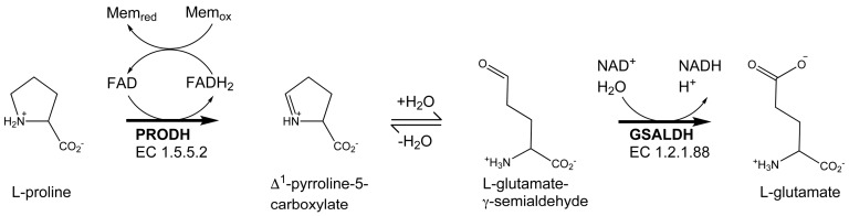
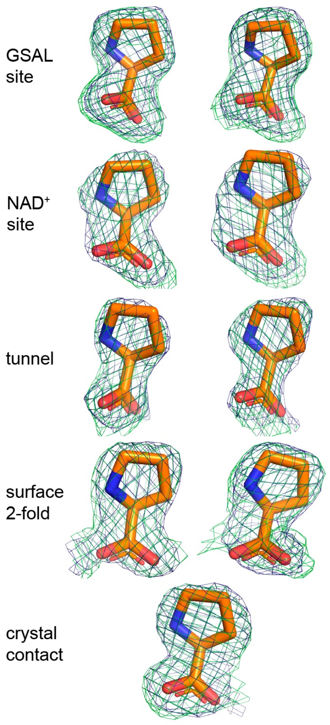
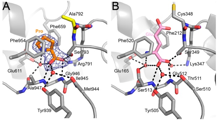
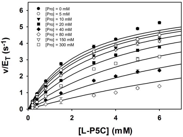
Proline utilization A (PutA) is a bifunctional flavoenzyme that catalyzes the two-step oxidation of l-proline to l-glutamate using spatially separated proline dehydrogenase (PRODH) and l-glutamate-γ-semialdehyde dehydrogenase (GSALDH) active sites. Substrate inhibition of the coupled PRODH-GSALDH reaction by proline is a common kinetic feature of PutAs, yet the structural basis for this phenomenon remains unknown. To understand the mechanism of substrate inhibition, we determined the 2.15 Å resolution crystal structure of Bradyrhizobium japonicum PutA complexed with proline. Proline was discovered in five locations remote from the PRODH active site. Most notably, strong electron density indicated that proline bound tightly to the GSAL binding site of the GSALDH active site. The pose and interactions of proline bound in this site are remarkably similar to those of the natural aldehyde substrate, GSAL, implying that proline inhibits the GSALDH reaction of PutA. Kinetic measurements show that proline is a competitive inhibitor of the PutA GSALDH reaction. Together, the structural and kinetic data show that substrate inhibition of the PutA coupled reaction is due to proline binding in the GSAL site.

Keywords: ">l-glutamate-γ-semialdehyde dehydrogenase; X-ray crystallography; flavoenzyme; proline dehydrogenase; substrate inhibition.


Conflict of interest statement

The authors declare no conflict of interest.

Figures

Figure 1
Figure 1
The reactions catalyzed by PutA.
Figure 2
Figure 2
The crystal structure of BjPutA C792A. (A) The protomer of BjPutA C792A. The PRODH module is colored blue. The GSALDH module is colored red. The purple surface represents the substrate-channeling tunnel. For reference, the two α-helices that border the central section of the tunnel are noted (α5a, 770 s helix). (B) Close-up view of proline molecules bound in the GSALDH active site and the middle of the tunnel. (C) The dimer of BjPutA C792A. The two protomers are colored gray and slate. The 9 proline molecules bound to the dimer are shown in spheres.
Figure 3
Figure 3
Electron density evidence showing the FAD of BjPutA C792A is reduced. (A) The FAD of proline-soaked BjPutA C792A. The mesh represents a simulated annealing FoFc omit map contoured at 3.0σ. The inset shows an edge-on view of the isoalloxazine. (B) The FAD of oxidized BjPutA (PDB ID 3HAZ).
Figure 4
Figure 4
Electron density of the 9 proline molecules bound to C792A. The green mesh represents a simulated annealing FoFc omit map contoured at 2.5σ. The blue mesh represents the refined 2FoFc map calculated from the final model, including proline ligands (1.0σ). The left and right sides of the figure show prolines bound to Chains A and B, respectively. Proline in the crystal contact does not have a non-crystallographic symmetry mate.
Figure 5
Figure 5
Proline inhibits the GSAL site of the GSALDH module. (A) Electron density and interactions for proline bound in the GSAL site. The mesh represents a simulated annealing FoFc omit map contoured at 3.0σ. (B) The active site of mouse GSALDH (ALDH4A1) complexed with the product glutamate (PDB ID 3V9K). Water molecules that mediate enzyme-ligand hydrogen bonds are represented by red spheres.
Figure 6
Figure 6
Kinetic data showing that proline is a competitive inhibitor of the GSALDH activity of BjPutA. Initial velocity pattern for BjPutA mutant R456M (0.25 μM) as a function of P5C concentration at 8 different fixed proline concentrations. The curves represent a global fit of the data to a competitive inhibition model. Best fit parameters were Km = 2.4 ± 0.1 mM, kcat = 6.7 ± 0.1 s−1, and Ki = 46.3 ± 1.7 mM proline.

References

    1. Liu L.K., Becker D.F., Tanner J.J. Structure, function, and mechanism of proline utilization a (puta) Arch. Biochem. Biophys. 2017;632:142–157. doi: 10.1016/j.abb.2017.07.005. - DOI - PMC - PubMed
    1. Tanner J.J. Structural biology of proline catabolic enzymes. Antioxid. Redox Signal. 2017 doi: 10.1089/ars.2017.7374. - DOI - PMC - PubMed
    1. Arentson B.W., Luo M., Pemberton T.A., Tanner J.J., Becker D.F. Kinetic and structural characterization of tunnel-perturbing mutants in bradyrhizobium japonicum proline utilization a. Biochemistry. 2014;53:5150–5161. doi: 10.1021/bi5007404. - DOI - PMC - PubMed
    1. Moxley M.A., Sanyal N., Krishnan N., Tanner J.J., Becker D.F. Evidence for hysteretic substrate channeling in the proline dehydrogenase and delta1-pyrroline-5-carboxylate dehydrogenase coupled reaction of proline utilization a (puta) J. Biol. Chem. 2014;289:3639–3651. doi: 10.1074/jbc.M113.523704. - DOI - PMC - PubMed
    1. Luo M., Gamage T.T., Arentson B.W., Schlasner K.N., Becker D.F., Tanner J.J. Structures of proline utilization a (puta) reveal the fold and functions of the aldehyde dehydrogenase superfamily domain of unknown function. J. Biol. Chem. 2016;291:24065–24075. doi: 10.1074/jbc.M116.756965. - DOI - PMC - PubMed

LinkOut - more resources