Review
. 2018 Jan 2;10(1):a018267.
doi: 10.1101/cshperspect.a018267.

The Actin Cytoskeleton and Actin-Based Motility

Affiliations
Review

The Actin Cytoskeleton and Actin-Based Motility

Tatyana Svitkina. Cold Spring Harb Perspect Biol. .

Abstract

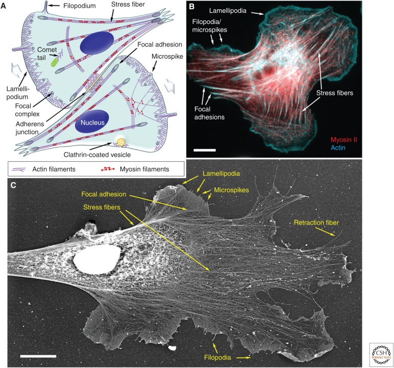
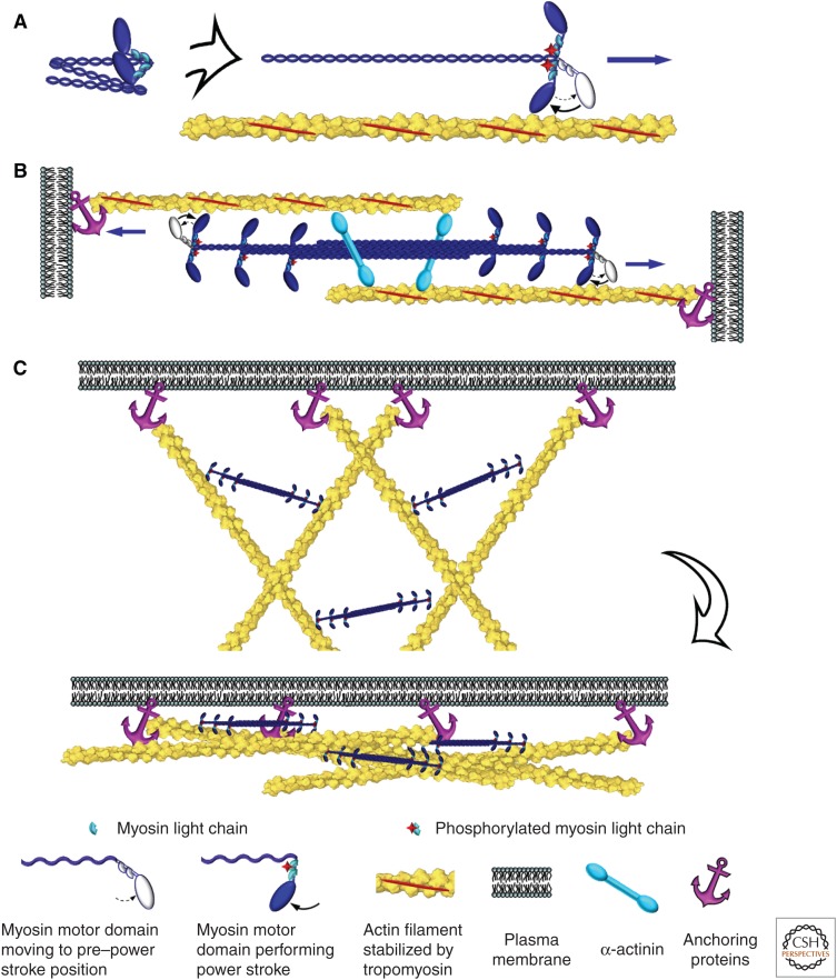
The actin cytoskeleton-a collection of actin filaments with their accessory and regulatory proteins-is the primary force-generating machinery in the cell. It can produce pushing (protrusive) forces through coordinated polymerization of multiple actin filaments or pulling (contractile) forces through sliding actin filaments along bipolar filaments of myosin II. Both force types are particularly important for whole-cell migration, but they also define and change the cell shape and mechanical properties of the cell surface, drive the intracellular motility and morphogenesis of membrane organelles, and allow cells to form adhesions with each other and with the extracellular matrix.


Figures

Figure 1.
Figure 1.
Components of the actin cytoskeleton in migrating cells. (A) Illustration of the components of the actin cytoskeleton in representative fibroblast-like cells. The direction of cell migration is indicated by wide gray arrows. (B) Fluorescence micrograph of a rat embryo fibroblast showing actin filaments (cyan) and myosin II (red). (C) Electron micrograph of the cytoskeleton of a Xenopus laevis fibroblast prepared by platinum shadowing after detergent extraction and critical point drying. Individual components of the actin cytoskeleton are marked in all panels. Scale bars, 10 µm. (C, Adapted from Svitkina and Borisy 1999.)
Figure 2.
Figure 2.
Accelerated dynamics of actin filaments. (A) Polymerization of the actin filament preferentially occurs at the barbed end from ATP-actin–profilin complexes; after incorporation of an actin monomer into the filament, profilin dissociates. Polymerization-triggered ATP hydrolysis and subsequent release of inorganic phosphate from actin subunits make filaments more susceptible to depolymerization and increase their affinity for actin-depolymerizing factor (ADF)/cofilin. ADF/cofilin severs filaments, promoting their depolymerization. Released actin subunits bind to profilin, which competes off ADF/cofilin and promotes nucleotide exchange in the actin monomer, thus producing new ATP-actin–profilin complexes. (B) Formins and (C) Ena/VASP (vasodilator-stimulated phosphoprotein) proteins associate with actin filament barbed ends, promote their elongation by recruiting actin–profilin complexes and protecting barbed ends from capping; they also anchor barbed ends to the membrane.
Figure 3.
Figure 3.
The organization of actin in filopodia and microspikes. (A) Platinum-replica electron microscopy of the cytoskeleton at the leading edge of a cultured mouse melanoma cell; actin filament bundles (cyan) that form the cytoskeleton of filopodia extend beyond the cell leading edge, span the dense actin filament network in lamellipodia (brown), and sometimes penetrate into the sparser actin network in the lamella (purple). Actin bundles in microspikes are mostly positioned within lamellipodia. Scale bar, 1 µm. (B) Enlarged boxed region from A shows long parallel actin filaments (cyan) and a complex of regulatory proteins (pink) at the filopodial tip. Some branched actin filaments in the adjacent lamellipodium are shaded orange. (C) Molecular organization of filopodia. Actin filaments are oriented with their barbed ends toward the filopodial tip. They are cross-linked into a bundle by fascin and laterally attached to the plasma membrane by ezrin-radixin-moesin (ERM) proteins. Formin and Ena/VASP (vasodilator-stimulated phosphoprotein) proteins associate with the actin filament barbed ends, anchor them to the membrane at the filopodial tip, and promote their elongation by protecting them from capping and recruiting actin–profilin complexes. Unconventional myosin X moves along actin filaments and accumulates at the filopodial tips. I-BAR-domain-containing proteins, such as IRSp53, help to maintain the tubular shape of the filopodial plasma membrane. Actin-depolymerizing factor (ADF)/cofilin and myosin II sever actin filaments at the filopodial base to stimulate actin filament depolymerization and monomer recycling.
Figure 4.
Figure 4.
Branched actin filament networks. (A) Organization of actin filaments in the lamellipodium of a fish epidermal keratocyte revealed by platinum-replica electron microscopy. A region outlined by the yellow box is enlarged in the yellow-framed inset to show branched actin filaments (highlighted in cyan). The red-framed inset shows the entire keratocyte moving upward. (B) Branched actin network in a comet tail assembled on ActA-coated latex beads in a cytoplasmic extract from Xenopus oocytes. A region outlined by the yellow box is enlarged in the yellow-framed inset to show branched actin filaments (highlighted in cyan) at the surface of the bead (pink). (C) Patches of branched actin filaments (cyan) assembled in the vicinity of clathrin-coated vesicles (yellow). (Reprinted from Collins et al. 2011.) (D) Cytoskeleton of an excitatory synapse in a cultured hippocampal neuron. A branched actin network in the dendritic spine (yellow) resides on dendritic microtubules (red) and forms a junction with the branched actin network in the apposing presynaptic bouton (green) associated with axonal microtubules (blue). The position of the junction is approximate. Scale bars, 500 nm (A,B,D); 100 nm (C); and 10 µm (red inset in A). (E) Diagram showing molecular organization of branched actin networks. The Arp2/3 complex is cooperatively activated by a membrane-targeted nucleation-promoting factor and a preexisting “mother” actin filament (1). On activation, the Arp2/3 complex nucleates a new “daughter” actin filament at the side of the mother filament and remains associated with the branchpoint; branchpoints are further stabilized by cortactin (2). The nascent filament elongates with its barbed end oriented toward the membrane. Its elongation is promoted by formins (3) and/or Ena/VASP proteins (4). After a period of elongation, the barbed end is capped by capping protein and lags behind the protruding leading edge (5). Disassembly at the rear of the lamellipodial network occurs through dissociation of branches and ADF/cofilin-mediated severing (6).
Figure 5.
Figure 5.
Contractile activity of nonmuscle myosin II. (A) A hexameric molecule of nonmuscle myosin II has a folded conformation in an inactive state (left). Phosphorylation of the myosin regulatory light chain causes unfolding of the myosin II molecule and restoration of motor activity; this activated myosin II can move toward the barbed end of the actin filament by swinging its motor domain in an ATP-dependent manner (right). Contraction of actin–myosin-II bundles (B) or networks (C) is mediated by bipolar filaments of myosin II that move along oppositely oriented (B) or nonaligned (C) actin filaments anchored at their barbed ends to the plasma membrane or other cellular structures.
Figure 6.
Figure 6.
Contractile actin structures in nonmuscle cells. (A) A stress fiber fragment (extends from upper left to lower middle) and a network of other cytoskeletal components (upper right half) in a rat fibroblast. Color coding: yellow, actin filaments; red, microtubules; cyan, intermediate filaments. (Reprinted from Svitkina et al. 1995.) (B) Immunogold labeling of a stress fiber (extends from upper left to lower right) with an antibody to nonmuscle myosin II. Distribution of colloidal gold particles shows semiperiodic arrangement of myosin II along the actin filament bundle. Color coding: yellow, gold particles; red, microtubules; cyan, intermediate filaments. (C) Dissolution of actin filaments by treatment of the cytoskeleton with the actin-severing protein gelsolin reveals stacks of bipolar myosin II filaments separated by irregular intervals. The diagonal alignment of myosin II filaments suggests that stacks belong to the same stress fiber running in an upper left to lower right direction. Color coding: brown, myosin II filaments; cyan, intermediate filaments; purple, clathrin-coated pits. (D) Contractile actin–myosin-II network in the lamella of a fish epidermal keratocyte. Immunogold staining of myosin II shows linear sets of gold particles (colored in yellow) representing myosin II filaments among the network of actin filaments. The orientation of actin and myosin II filaments changes from the front (right) to the rear (left) of the cell, reflecting network contraction during keratocyte migration. (E) A cluster of nonoriented bipolar filaments of nonmuscle myosin II in the lamella of a fish epidermal keratocyte visualized after dissolution of actin filaments by gelsolin treatment. Filaments form chains (upper right), networks (center), and stacks (lower left). Several individual bipolar filaments are colored in brown. Scale bars, 500 nm (AC) and 250 nm (D,E).

References

    1. Abercrombie M, Heaysman JE, Pegrum SM. 1970. The locomotion of fibroblasts in culture. II. ‘Ruffling’. Exp Cell Res 60: 437–444. - PubMed
    1. Abmayr SM, Pavlath GK. 2012. Myoblast fusion: Lessons from flies and mice. Development 139: 641–656. - PMC - PubMed
    1. Adelstein RS, Conti MA. 1975. Phosphorylation of platelet myosin increases actin-activated myosin ATPase activity. Nature 256: 597–598. - PubMed
    1. Alexandrova AY, Arnold K, Schaub S, Vasiliev JM, Meister JJ, Bershadsky AD, Verkhovsky AB. 2008. Comparative dynamics of retrograde actin flow and focal adhesions: Formation of nascent adhesions triggers transition from fast to slow flow. PLoS ONE 3: e3234. - PMC - PubMed
    1. Beach JR, Shao L, Remmert K, Li D, Betzig E, Hammer JA. 3rd 2014. Nonmuscle myosin II isoforms coassemble in living cells. Curr Biol 24: 1160–1166. - PMC - PubMed

Publication types

LinkOut - more resources