. 2018 Jan 2;9(1):35.
doi: 10.1038/s41467-017-02441-z.

Extreme sensitivity to ultraviolet light in the fungal pathogen causing white-nose syndrome of bats

Affiliations

Extreme sensitivity to ultraviolet light in the fungal pathogen causing white-nose syndrome of bats

Jonathan M Palmer et al. Nat Commun. .

Abstract

Bat white-nose syndrome (WNS), caused by the fungal pathogen Pseudogymnoascus destructans, has decimated North American hibernating bats since its emergence in 2006. Here, we utilize comparative genomics to examine the evolutionary history of this pathogen in comparison to six closely related nonpathogenic species. P. destructans displays a large reduction in carbohydrate-utilizing enzymes (CAZymes) and in the predicted secretome (~50%), and an increase in lineage-specific genes. The pathogen has lost a key enzyme, UVE1, in the alternate excision repair (AER) pathway, which is known to contribute to repair of DNA lesions induced by ultraviolet (UV) light. Consistent with a nonfunctional AER pathway, P. destructans is extremely sensitive to UV light, as well as the DNA alkylating agent methyl methanesulfonate (MMS). The differential susceptibility of P. destructans to UV light in comparison to other hibernacula-inhabiting fungi represents a potential "Achilles' heel" of P. destructans that might be exploited for treatment of bats with WNS.


Conflict of interest statement

The authors declare no competing financial interests.

Figures

Fig. 1
Fig. 1
Maximum likelihood phylogeny of Pseudogymnoascus draft genomes and several outgroup Ascomycetes calibrated using fossil evidence. The six nonpathogenic Pseudogymnoascus species sequenced here are among the closest known relatives of P. destructans. P. destructans was estimated to have diverged from its last common ancestor around 23.5 MYA. Node support values are derived from 1000 bootstrap replicates
Fig. 2
Fig. 2
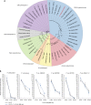
Comparative genomic analyses of orthologous proteins of Pseudogymnoascus destructans and six close relatives. Maximum likelihood phylogeny illustrates relationships between fungi used in this study. Genome-level comparisons made based on number of gene models, number of CAZymes, number of proteases, and the number of secreted proteins as described in Methods
Fig. 3
Fig. 3
Heatmap of Biolog phenotypic microarrays testing fungal growth. Carbon utilization from 190 sources was tested to assess the growth of each of seven Pseudogymnoascus species. Growth was quantified using ImageJ ColonyArea plug-in after 15 °C incubation for 7 days for the nonpathogenic Pseudogymnoascus species and after 14 days for P. destructans. Growth for each carbon source is presented as normalized to growth on glucose. These data are derived from a single biological replicate (n = 1)
Fig. 4
Fig. 4
Sensitivity of Pseudogymnoascus species to DNA-damaging agents. a Qualitative plate assay measuring the effects of four DNA-damaging agents on the growth of each fungal species (4-nitroquinoline (4-NQO), camptothecin (CPT), methyl methanesulfonate (MMS), and 254-nm ultraviolet light (UV-C)). Fungal spores were serially diluted, inoculated on appropriate medium, and growth was quantified after 7-day incubation at 15 °C. Multiple isolates of P. destructans were tested alongside the nonpathogenic Pseudogymnoascus species. b A quantitative assay using colony-forming units (CFUs) to measure survival of each fungus under different wavelengths of UV light (254 nm (UV-C), 312 nm (UV-B), and 366 nm (UV-A)). CFU assays were conducted in biological triplicate (n = 3) and error bars represent standard deviations
Fig. 5
Fig. 5
Blue light-mediated photoreactivation DNA repair is not functional in P. destructans. a Characterization of photolyases in Pseudogymnoascus species based on protein alignment and maximum likelihood phylogeny of well-characterized enzymes. Pseudogymnoascus destructans lacks a canonical CPD photolyase I, although it harbors a cyr-DASH photolyase (VC83_00225). The genomes of Pseudogymnoascus verrucosus, P. sp. 24MN13, and P. sp. 05NY08 contain a CPD photolyase I, whereas all species studied contain at least one cry-DASH ortholog. Proteins identified in ref. were used for phylogenetic comparison of DNA photolyases. b Photoreactivation experiments comparing survival of germinating conidia exposed to varying doses of UV-C (254 nm) followed by treatment with UV-A (366 nm) for 1 h indicate that only P. verrucosus and P. sp. 24MN13 display increased survival attributed to the activity of CPD photolyase I. No change in survival was detected in P. destructans or the other three nonpathogenic Pseudogymnoascus species tested. Interestingly, P. sp. 05NY08 did not show an increase in CFU survival despite the fungus harboring a CPD photolyase I enzyme. A similar phenomenon occurs in Aspergillus nidulans, where there was no change in CFU survival in photoreactivation assays in wild-type background, despite the presence of the functional CryA gene (CPD photolyase I). CFU assays were performed in biological triplicate (n = 3) and error bars represent standard deviation

Similar articles

Cited by

References

    1. Lorch JM, et al. First detection of bat white-nose syndrome in western North America. mSphere. 2016;1:e00148–16. doi: 10.1128/mSphere.00148-16. - DOI - PMC - PubMed
    1. Frick WF, et al. An emerging disease causes regional population collapse of a common North American bat species. Science. 2010;329:679–682. doi: 10.1126/science.1188594. - DOI - PubMed
    1. Gargas A, Trest MT, Christensen M, Volk TJ, Blehert DS. Geomyces destructans sp. nov. associated with bat white-nose syndrome. Mycotaxon. 2009;108:147–154. doi: 10.5248/108.147. - DOI
    1. Minnis AM, Lindner DL. Phylogenetic evaluation of Geomyces and allies reveals no close relatives of Pseudogymnoascus destructans, comb. nov., in bat hibernacula of eastern North America. Fungal Biol. 2013;117:638–649. doi: 10.1016/j.funbio.2013.07.001. - DOI - PubMed
    1. Verant ML, Boyles JG, Waldrep W, Wibbelt G, Blehert DS. Temperature-dependent growth of Geomyces destructans, the fungus that causes bat white-nose syndrome. PLoS ONE. 2012;7:e46280. doi: 10.1371/journal.pone.0046280. - DOI - PMC - PubMed

Publication types