

Establishment and evaluation of four different types of patient-derived xenograft models
- PMID: 29296105
- PMCID: PMC5738885
- DOI: 10.1186/s12935-017-0497-4
Establishment and evaluation of four different types of patient-derived xenograft models
Abstract
Background: Patient-derived xenografts (PDX) have a biologically stable in tumor architecture, drug responsiveness, mutational status and global gene-expression patterns. Numerous PDX models have been established to date, however their thorough characterization regarding the tumor formation and rates of tumor growth in the established models remains a challenging task. Our study aimed to provide more detailed information for establishing the PDX models successfully and effectively.
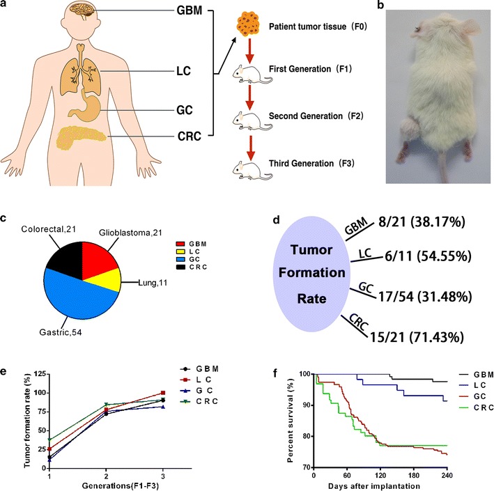
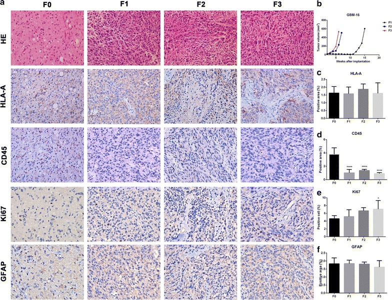
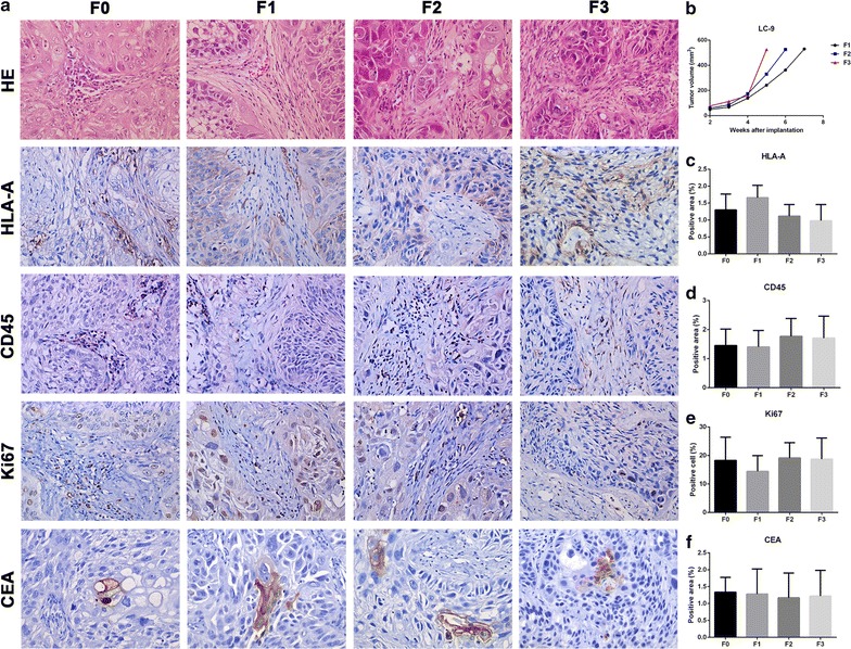
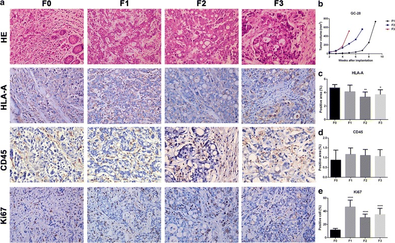
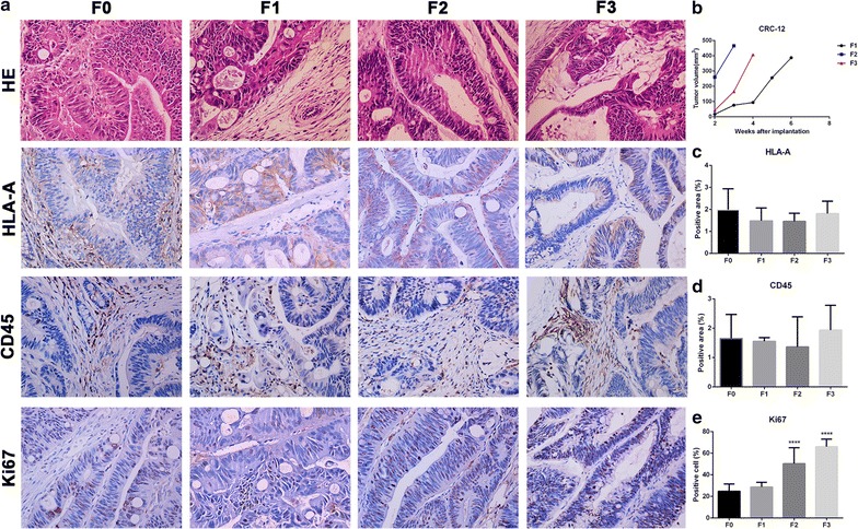
Methods: We transplanted four different types of solid tumors from 108 Chinese patients, including 21 glioblastoma (GBM), 11 lung cancers (LC), 54 gastric cancers (GC) and 21 colorectal cancers (CRC), and took tumor tissues passaged for three successive generations. Here we report the rate of tumor formation, tumor-forming times, tumor growth curves and mortality of mice in PDX model. We also report H&E staining and immunohistochemistry for HLA-A, CD45, Ki67, GFAP, and CEA protein expression between patient cancer tissues and PDX models.
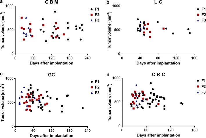
Results: Tumor formation rate increased significantly in subsequent tumor generations. Also, the survival rates of GC and CRC were remarkably higher than GBM and LC. As for the time required for the formation of tumors, which reflects the tumor growth rate, indicated that tumor growth rate always increased as the generation number increased. The tumor growth curves also illustrate this law. Similarly, the survival rate of PDX mice gradually improved with the increased generation number in GC and CRC. And generally, there was more proliferation (Ki67+) in the PDX models than in the patient tumors, which was in accordance with the results of tumor growth rate. The histological findings confirm similar histological architecture and degrees of differentiation between patient cancer tissues and PDX models with statistical analysis by GraphPad Prism 5.0.
Conclusion: We established four different types of PDX models successfully, and our results add to the current understanding of the establishment of PDX models and may contribute to the extension of application of different types of PDX models.
Keywords: Colorectal cancer (CRC); Gastric cancer (GC); Glioblastoma (GBM); Lung cancer (LC); Patient derived xenograft (PDX).
Figures
Similar articles
-
Establishment of Novel Gastric Cancer Patient-Derived Xenografts and Cell Lines: Pathological Comparison between Primary Tumor, Patient-Derived, and Cell-Line Derived Xenografts.Cells. 2019 Jun 14;8(6):585. doi: 10.3390/cells8060585. Cells. 2019. PMID: 31207870 Free PMC article.
-
An improved method to build lung cancer PDX models by surgical resection samples and its association with B7-H3 expression.Transl Cancer Res. 2019 Dec;8(8):2848-2857. doi: 10.21037/tcr.2019.10.40. Transl Cancer Res. 2019. PMID: 35117042 Free PMC article.
-
Patient-derived xenograft models for gastrointestinal tumors: A single-center retrospective study.Front Oncol. 2022 Nov 18;12:985154. doi: 10.3389/fonc.2022.985154. eCollection 2022. Front Oncol. 2022. PMID: 36465411 Free PMC article.
-
Current Update of Patient-Derived Xenograft Model for Translational Breast Cancer Research.J Mammary Gland Biol Neoplasia. 2017 Jun;22(2):131-139. doi: 10.1007/s10911-017-9378-7. Epub 2017 Apr 27. J Mammary Gland Biol Neoplasia. 2017. PMID: 28451789 Free PMC article. Review.
-
Applications of patient-derived tumor xenograft models and tumor organoids.J Hematol Oncol. 2020 Jan 7;13(1):4. doi: 10.1186/s13045-019-0829-z. J Hematol Oncol. 2020. PMID: 31910904 Free PMC article. Review.
Cited by
-
Targeted therapy of the AKT kinase inhibits esophageal squamous cell carcinoma growth in vitro and in vivo.Int J Cancer. 2019 Aug 15;145(4):1007-1019. doi: 10.1002/ijc.32285. Epub 2019 Apr 3. Int J Cancer. 2019. PMID: 30887517 Free PMC article.
-
ROS-mediated inactivation of the PI3K/AKT pathway is involved in the antigastric cancer effects of thioredoxin reductase-1 inhibitor chaetocin.Cell Death Dis. 2019 Oct 24;10(11):809. doi: 10.1038/s41419-019-2035-x. Cell Death Dis. 2019. PMID: 31649256 Free PMC article.
-
Direct Implantation of Patient Brain Tumor Cells into Matching Locations in Mouse Brains for Patient-Derived Orthotopic Xenograft Model Development.Cancers (Basel). 2024 Apr 28;16(9):1716. doi: 10.3390/cancers16091716. Cancers (Basel). 2024. PMID: 38730671 Free PMC article.
-
Advanced Xenograft Model with Cotransplantation of Patient-Derived Organoids and Endothelial Colony-Forming Cells for Precision Medicine.J Oncol. 2021 Jul 13;2021:9994535. doi: 10.1155/2021/9994535. eCollection 2021. J Oncol. 2021. PMID: 34335765 Free PMC article.
-
Modeling Glioma Stem Cell-Mediated Tumorigenesis Using Zebrafish Patient-Derived Xenograft Systems.Methods Mol Biol. 2025;2944:257-277. doi: 10.1007/978-1-0716-4654-0_20. Methods Mol Biol. 2025. PMID: 40553289
References
LinkOut - more resources
Full Text Sources
Other Literature Sources
Research Materials
Miscellaneous
