. 2017 Nov 18;8(64):107477-107491.
doi: 10.18632/oncotarget.22491. eCollection 2017 Dec 8.

Interleukin enhancer-binding factor 3 and HOXC8 co-activate cadherin 11 transcription to promote breast cancer cells proliferation and migration

Affiliations

Interleukin enhancer-binding factor 3 and HOXC8 co-activate cadherin 11 transcription to promote breast cancer cells proliferation and migration

Yang Zhang et al. Oncotarget. .

Abstract

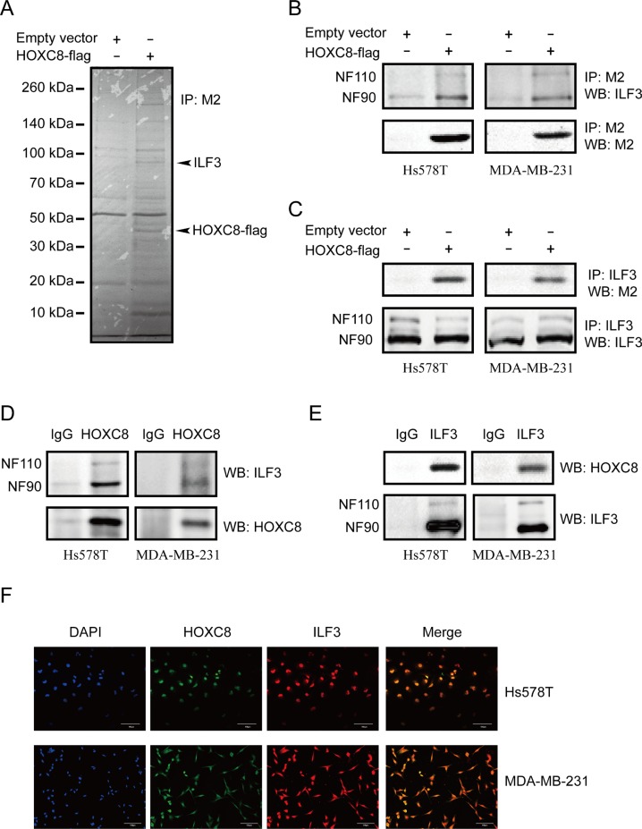
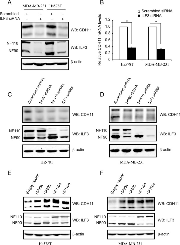
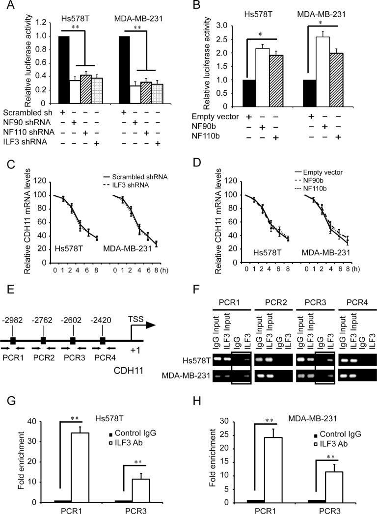
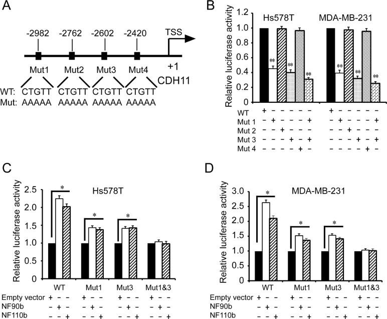
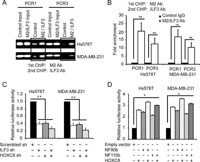
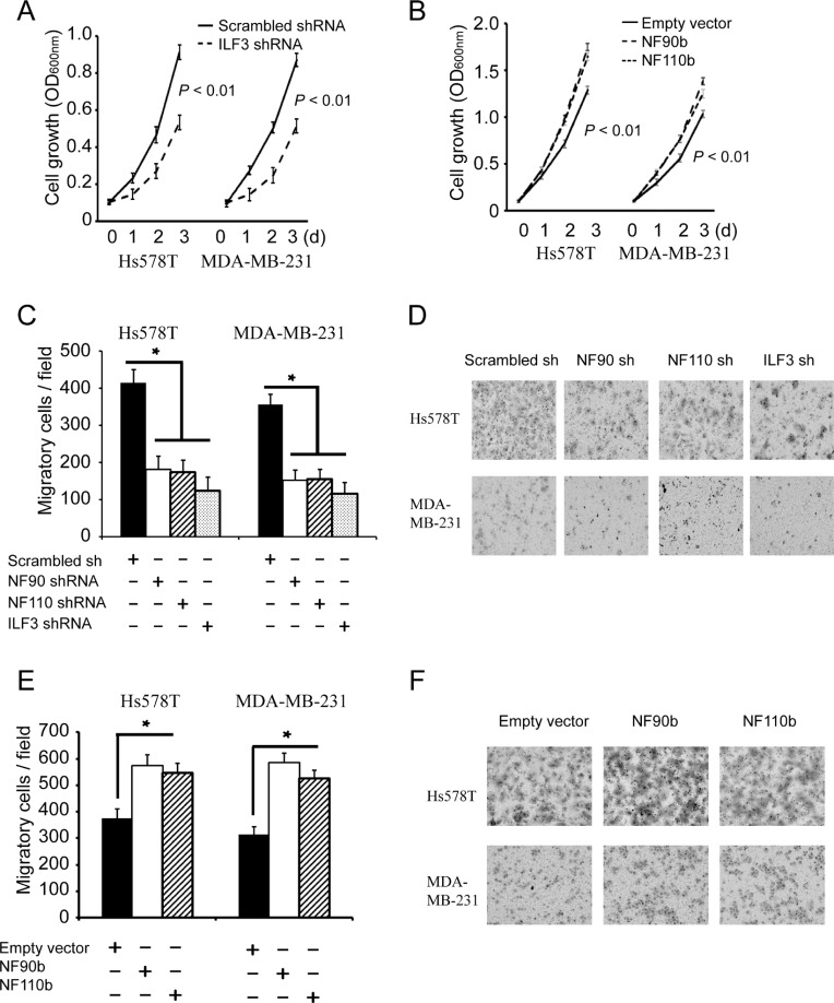
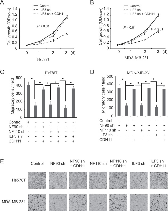
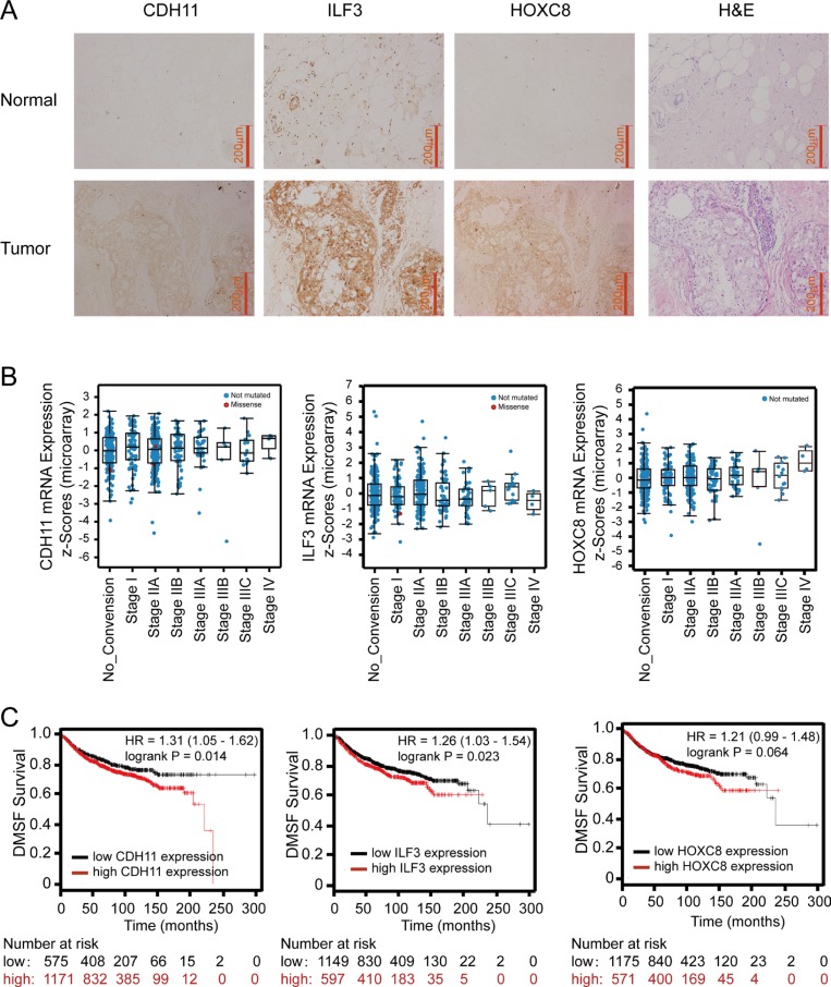
Cadherin 11 (CDH11) expression is detected only in invasive breast cancer cells and aggressive breast cancer specimens. However, little is known about the molecular mechanisms of CDH11 transcriptional regulation. Here, we report that interleukin enhancer binding factor 3 (ILF3) interacts with Homeobox C8 (HOXC8) to activate CDH11 transcription in breast cancer cells. Using co-immunoprecipitation and mass spectrometry analyses, ILF3 is shown to interact with HOXC8 in breast cancer cells. We demonstrate that ILF3 binds to the CDH11 promoter on nucleotides -2982 ~ -2978 and -2602 ~ 2598 and interacts with HOXC8 to co-activate CDH11 transcription. We further show that ILF3 promotes proliferation and migration, at least partially, by facilitating CDH11 expression in breast cancer cells. Moreover, immunohistochemistry (IHC) shows that expression of CDH11, ILF3 and HOXC8 are all upregulated in breast cancer specimens compared to normal breast tissues. Importantly, the expression levels of CDH11, ILF3 and HOXC8 are elevated in the advanced stages of breast cancer, and high expression of CDH11, ILF3 and HOXC8 is associated with poor distant metastasis-free survival (DMFS) for breast cancer patients.

Keywords: CDH11; HOXC8; ILF3; breast cancer; transcriptional regulation.


Conflict of interest statement

CONFLICTS OF INTEREST The authors declare that they have no competing interest.

Figures

Figure 1
Figure 1. ILF3 interacts with HOXC8 in breast cancer cells
(A) MDA-MB-231 cells were lentivirally transduced with the empty vector or vector encoding HOXC8-flag. Co-immunoprecipitations performed with the anti-flag M2 antibody were separated by SDS–PAGE, and the differential bands were analyzed by MS. (B) Co-immunoprecipitation (Co-IP) was performed using the M2 antibody in Hs578T or MDA-MB-231 cells that were lentivirally transduced with the empty vector or vector encoding HOXC8-flag. Immunoprecipitates were analyzed by Western blotting (WB). (C) Co-IP was performed using the ILF3 antibody in Hs578T or MDA-MB-231 cells that were lentivirally transduced with the empty vector or vector encoding HOXC8-flag. Immunoprecipitates were analyzed by WB. (D) Co-IP experiment was performed using anti-HOXC8 antibody in Hs578T or MDA-MB-231 cells, and precipitated proteins were analyzed by WB using anti-ILF3 or anti-HOXC8 antibodies as indicated. (E) Co-IP experiment was performed using anti-ILF3 antibody in Hs578T or MDA-MB-231 cells, and precipitated proteins were analyzed by WB using anti-HOXC8 or ILF3 antibodies as indicated. (F) Immunofluorescence staining for HOXC8-flag (green) and ILF3 (red) in Hs578T or MDA-MB-231 cells that were lentivirally transduced with the vector encoding HOXC8-flag; DAPI staining for the nucleus. Magnification, 200×; Scale bar, 100 μm.
Figure 2
Figure 2. ILF3 activates CDH11 expression in the Hs578T and MDA-MB-231 cell lines
(A) Hs578T and MDA-MB-231 cells were transfected with siRNAs that specifically targeted ILF3 or with scrambled siRNAs. Cell lysates were immunoblotted for CDH11, ILF3 and β-actin, as indicated. (B) Hs578T and MDA-MB-231 cells were transfected with siRNAs that specifically targeted ILF3 or with scrambled siRNAs. Total RNA was subjected to qRT-PCR to examine the levels of CDH11 mRNA. β-actin mRNA was used as an internal control for standardization. (C and D) Hs578T or MDA-MB-231 cells were lentivirally transduced with NF90-, NF110- or ILF3-specific shRNA, and Western blotting was performed with antibodies for CDH11, ILF3 or β-actin, as indicated. (E and F) Hs578T or MDA-MB-231 cells were lentivirally transduced with NF90a, NF90b, NF110a or NF110b expression vectors, and Western blotting was performed with antibodies for CDH11, ILF3 or β-actin, as indicated.
Figure 3
Figure 3. ILF3 binds to CDH11 promoter and activates CDH11 transcription in breast cancer cells
(A) Luciferase assays were performed in cells transduced with CDH11 promoter luciferase reporter vectors in NF90-, NF110- or ILF3-specific shRNA knockdown cells. The luciferase activity was normalized to the Renilla activity. (B) Luciferase assays were performed in cells transduced with CDH11 promoter luciferase reporter vectors in NF90b or NF110b ecto-expression cells. (C) Control or ILF3-knockdown cells were treated with 1 μg/ml actinomycin D for varying times (0, 1, 2, 4, 6 and 8 h). Total RNA was extracted and then subjected to qRT-PCR to analyze the level of CDH11 mRNA. β-actin mRNA was used as the internal standard. (D) Cells transduced with vectors encoding NF90b, NF110b or empty vectors were treated with 1μg/ml actinomycin D for varying times (0, 1, 2, 4, 6 and 8 h). Total RNA was extracted and then subjected to qRT-PCR to analyze the level of CDH11 mRNA. The level of β-actin mRNA was used as the internal standard. (E) schematic diagram of the positions of the putative ILF3 binding sites on the CDH11 promoter. Arrows show the regions for PCR primer amplification. (F) ChIP was performed using the ILF3 antibody, and the immunoprecipitated chromatin DNA was subjected to PCR. (G and H) ChIP was performed using the ILF3 antibody, and the immunoprecipitated chromatin DNA was subjected to real-time PCR. Each sample was run in triplicate and in multiple experiments for mean ± SEM; *P < 0.05; **P < 0.01.
Figure 4
Figure 4. Identification of the ILF3 binding sites in the CDH11 promoter by mutagenesis
(A) A schematic diagram for the mutagenesis of four CTGTT (putative ILF3 binding) sites in the CDH11 promoter. (B) Luciferase analyses were performed with wild-type (WT) or mutant (Mut) CDH11 promoter luciferase reporter vectors in Hs578T or MDA-MB-231 cells. The luciferase activity was measured and normalized to the Renilla activity. Columns, mean; bars, SEM; **P < 0.01. (C and D) Hs578T or MDA-MB-231 cells were lentivirally transduced with NF90b, NF110b expression vectors or the empty vectors and then transfected with mutant (Mut) CDH11 promoter luciferase reporter vectors, as indicated. The luciferase activity was measured and normalized to the Renilla activity. Each sample was run in triplicate and in multiple experiments for mean ± SEM; *P < 0.05; **P < 0.01.
Figure 5
Figure 5. ILF3 and HOXC8 co-occupy the CDH11 promoter to activate CDH11 transcription
(A and B) Sequential ChIP assays were performed in Hs578T or MDA-MB-231 cells that were lentivirally transduced with HOXC8-flag expression vectors. Chromatin was incubated with the anti-flag M2 antibody, and the immunocomplexes were subjected to a second round of ChIP using antibodies against ILF3 or with rabbit IgG as the control. Precipitated chromatin DNA was subjected to PCR or real-time PCR. (C) Cells were lentivirally transduced with ILF3 shRNA, HOXC8 shRNA or scrambled shRNA vectors as indicated and transfected with CDH11 promoter luciferase reporter plasmids. The luciferase activity was measured and normalized to the Renilla activity. (D) Cells were lentivirally transduced with vectors encoding NF90b, NF110b or HOXC8 and transfected with CDH11 promoter luciferase reporter plasmids. The luciferase activity was measured and normalized to the Renilla activity. Each sample was run in triplicate and in multiple experiments for mean ± SEM; *P < 0.05; **P < 0.01.
Figure 6
Figure 6. ILF3 promotes proliferation and migration breast cancer cells
(A) MTT assays to analyze cell proliferation of control, or ILF3 knockdown cells. Data are the mean ± SEM; n = 3; P < 0.01. (B) MTT assays were carried out in cells that were lentivirally transduced with empty vectors or vectors encoding NF90b or NF110b. Data are the mean ± SEM; n = 3; P < 0.01. (C and D) Transwell assays to analyze cell migration of control, NF90, NF110 or ILF3 knockdown cells. Columns, mean; bars, SEM; n = 3; *P < 0.05. (E and F) Transwell assays to analyze migration of cells that were lentivirally transduced with empty vectors or vectors encoding NF90b or NF110b. Columns, mean; bars, n = 3; SEM; *P < 0.05.
Figure 7
Figure 7. The effects of ILF3 knockdown on cell proliferation and migration are rescued by ecto-expression of CDH11 in breast cancer cell lines
(A and B) ILF3 knockdown cells were lentivirally transduced with CDH11 expression vectors, and MTT assays were performed to analyze cell proliferation. Data are the mean ± SEM; n = 3; P < 0.01. (C and D) Hs578T or MDA-MB-231 cells that were transduced with NF90 shRNA, NF110 shRNA or ILF3 shRNA were lentivirally transduced with CDH11 expression vectors, and Transwell assays were performed to analyze cell migration. Columns, mean; bars, SEM; n = 3; *P < 0.05. (E) Representative images of migratory cells that are crystal violet-stained on the undersurface of transwell.
Figure 8
Figure 8. Immunohistochemistry of ILF3, CDH11 and HOXC8 in breast cancer specimens and analyses of ILF3, CDH11 and HOXC8 expression using publicly available datasets
(A) IHC staining of CDH11, ILF3 and HOXC8 in normal breast and breast cancer tissues. Magnification, 200×; Scale bar, 200 μm. (B) mRNA expression levels of CDH11, ILF3 and HOXC8 in breast cancer specimens from different disease stages using the breast cancer dataset (Breast invasive carcinoma, TCGA, Nature 2012, 825 samples) from cBioPortal (www.cbioportal.org). (C) Kaplan-Meier survival plots show distant metastasis-free survival for breast cancer patients with high and low expression of CDH11, ILF3 or HOXC8. Data were analyzed using an online survival analysis tool (www.kmplot.com, probe ID: 217804_s_at for ILF3, 207173_x_at for CDH11 and 221350_at for HOXC8).

References

    1. Thiery JP. Epithelial-mesenchymal transitions in tumour progression. Nat Rev Cancer. 2002;2:442–454. - PubMed
    1. Hazan RB, Qiao R, Keren R, Badano I, Suyama K. Cadherin switch in tumor progression. Ann N Y Acad Sci. 2004;1014:155–163. - PubMed
    1. Okazaki M, Takeshita S, Kawai S, Kikuno R, Tsujimura A, Kudo A, Amann E. Molecular cloning and characterization of OB-cadherin, a new member of cadherin family expressed in osteoblasts. J Biol Chem. 1994;269:12092–12098. - PubMed
    1. Simonneau L, Kitagawa M, Suzuki S, Thiery JP. Cadherin 11 expression marks the mesenchymal phenotype: towards new functions for cadherins? Cell Adhes Commun. 1995;3:115–130. - PubMed
    1. Pishvaian MJ, Feltes CM, Thompson P, Bussemakers MJ, Schalken JA, Byers SW. Cadherin-11 is expressed in invasive breast cancer cell lines. Cancer Res. 1999;59:947–952. - PubMed