. 2017 Sep 21;7(1):e1373235.
doi: 10.1080/2162402X.2017.1373235. eCollection 2017.

The "don't eat me" signal CD47 is a novel diagnostic biomarker and potential therapeutic target for diffuse malignant mesothelioma

Affiliations

The "don't eat me" signal CD47 is a novel diagnostic biomarker and potential therapeutic target for diffuse malignant mesothelioma

Christian M Schürch et al. Oncoimmunology. .

Abstract

Diffuse malignant mesothelioma (DMM) is one of the prognostically most discouraging cancers with median survivals of only 12-22 months. Due to its insidious onset and delayed detection, DMM is often at an advanced stage at diagnosis and is considered incurable. Combined chemo- and radiotherapy followed by surgery only marginally affect outcome at the cost of significant morbidity. Because of the long time period between exposure to asbestos and disease onset, the incidence of DMM is still rising and predicted to peak around 2020. Novel markers for the reliable diagnosis of DMM in body cavity effusion specimens as well as more effective, targeted therapies are urgently needed. Here, we show that the "don't eat me" signalling molecule CD47, which inhibits phagocytosis by binding to signal regulatory protein α on macrophages, is overexpressed in DMM cells. A two-marker panel of high CD47 expression and BRCA1-associated protein 1 (BAP-1) deficiency had a sensitivity of 78% and specificity of 100% in discriminating DMM tumour cells from reactive mesothelial cells in effusions, which is superior to the currently used four-marker combination of BAP-1, glucose transporter type 1, epithelial membrane antigen and desmin. In addition, blocking CD47 inhibited growth and promoted phagocytosis of DMM cell lines by macrophages in vitro. Furthermore, DMM tumours in surgical specimens from patients as well as in a mouse DMM model expressed high levels of CD47 and were heavily infiltrated by macrophages. Our study demonstrates that CD47 is an accurate novel diagnostic DMM biomarker and that blocking CD47 may represent a promising therapeutic strategy for DMM.

Keywords: CD47; biomarker; calreticulin; cancer; diffuse malignant mesothelioma; macrophages; phagocytosis; targeted therapy; “don't eat me” signal.


Figures

Figure 1.
Figure 1.
The “don't eat me” signal CD47 and the pro-phagocytic molecule calreticulin are expressed in diffuse malignant mesothelioma. (A) Relative expression of CD47, CALR, MUC1 (EMA), DES, SLC2A1 (GLUT1) and BAP1 mRNA in normal pleura (NP; n = 5) vs. pleural DMM (n = 40) were analysed in a publicly available microarray dataset (GSE2549) using the Gene Expression Omnibus GEO2R online tool. (B) CD47 and calreticulin expression on mesothelioma cell lines ACC-MESO-1 and H28, respectively, as analysed by FACS. One representative histogram of 9 (ACC-MESO-1) and 4 (H28) per staining is shown. Red lines, CD47 and calreticulin stainings; blue lines, respective isotype controls. (C) Mean fluorescence intensities (MFI) of CD47 vs. isotype (left panel) and calreticulin vs. isotype (right panel) in 5 different DMM cell lines. (D) Immunohistochemistry for CD47 on FFPE cell blocks from DMM cell lines. Examples of low (1+) and high (3+) CD47 expression are shown. Scale bars, 50μm. Statistics: (A) Mann-Whitney test; (C) paired t-test. *p < 0.05; ****p < 0.0001
Figure 2.
Figure 2.
CD47 immunohistochemistry discriminates DMM from reactive mesothelial cells in effusion cytology. (A-B) Representative examples of different CD47 immunohistochemistry staining intensities in DMM and reactive effusions in TMAs constructed of cell blocks from effusion specimens. Each row represents one patient. The corresponding stainings for calretinin, EMA, BAP-1, desmin, GLUT-1, calreticulin and H&E morphology are shown. Staining intensity for the respective molecules was assessed in mesothelial cells identified using calretinin. The interpretation of the result is indicated at the upper right of each spot. (A) Diffuse malignant mesothelioma, CD47 staining intensity low (0) and high (2+, 3+). (B) Reactive effusion, CD47 staining intensity low (0). Scale bars, 100μm; original magnification 150x. Staining assessment: CD47 (membranous staining): 0, negative; 1+, incomplete or weak; 2+, complete, moderately strong; 3+, complete, strong. EMA (membranous staining, modified according to 5): lo, negative or 1+ and <25% positive cells; hi, 2+ or 3+ or 4+ and ≥25% positive cells. BAP-1 (nuclear staining): L, lost; R, retained. Desmin (cytoplasmic staining): -, loss in any number of mesothelial cells; +, no loss. GLUT-1 (membranous staining): -, negative; +, positive. Calreticulin (cytoplasmic and membranous staining): lo, <95% positive cells; hi, ≥95% positive cells.
Figure 3.
Figure 3.
Blocking CD47 inhibits cell growth and promotes phagocytosis of malignant mesothelioma cell lines. (A) DMM cell lines were cultured in duplicates for three days in the presence of 5μg/ml isotype control (IgG) or anti-CD47 (αCD47) mAb and cell numbers were assessed daily in a Neubauer chamber using trypan blue staining. The fold increase in cell growth of IgG- vs. αCD47-treated DMM cell lines is shown for each day. Pooled data from 5 independent experiments. (B) Schematic of phagocytosis assay. Differentiated macrophages were serum starved for 2h and then co-cultured with CFSE-labelled ACC-MESO-1 cells for 2h in the presence of 10μg/ml IgG or αCD47 mAb, followed by FACS analysis. (C) Representative dot plots of CD45+CD14+ macrophages containing CFSE+ ACC-MESO-1 cells. Non-phagocytized CFSE+ tumour cells are gated in the left panels (top left). (D) Normalized fold increase in phagocytosis of αCD47 vs. IgG mAb-treated co-cultures (IgG = 1). Pooled data from two independent experiments run in duplicates or triplicates are shown. Results (mean±SEM), experiment 1: IgG, 18.7% ± 2.45%; αCD47 34.78% ± 4.28%; experiment 2: IgG 0.73% ± 0.12%; αCD47 1.15% ± 0.08%. f.i., fold increase. Statistics: (A, D) one-sample t-test (hypothetical value = 1). *p < 0.05; **p < 0.01.
Figure 4.
Figure 4.
Surgical DMM specimens express CD47 and are heavily infiltrated by macrophages. Representative examples of CD47 high (Patient 2) and low (Patient 3) expressing pleural effusions and matched surgical resection specimens are shown. (A) CD47 immunohistochemistry of the effusion cell block (cf. Fig. 2). (B-E) H&E staining (B) and immunohistochemistry for CD47 (C), CD68 (D) and CD163 (E). Scale bars, 40μm; original magnification 400x.
Figure 5.
Figure 5.
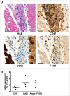
CD47 is expressed in a murine model of DMM. A) H&E staining and immunohistochemistry for CD47, CD68 and F4/80 in subcutaneously transplanted primary abdominal murine mesothelioma tumours. One representative example of n = 5 mice is shown. (B) Expression levels of Cd47, Cd68 and Adgre (F4/80) mRNA in murine tumours from (A). Scale bars, 50μm; original magnification 400x.

References

    1. Moolgavkar S, Chang E, G M, FS M. Epidemiology of Mesothelioma. In: Testa J, ed. Current Cancer Research – Asbestos and Mesothelioma. Cham, Switzerland: Springer, 2017.
    1. Alley EW, Katz SI, Cengel KA, Simone CB, 2nd. Immunotherapy and radiation therapy for malignant pleural mesothelioma. Transl Lung Cancer Res. 2017;6:212-9. doi:10.21037/tlcr.2017.04.01. PMID:28529903 - DOI - PMC - PubMed
    1. Hasteh F, Lin GY, Weidner N, Michael CW. The use of immunohistochemistry to distinguish reactive mesothelial cells from malignant mesothelioma in cytologic effusions. Cancer Cytopathol. 2010;118:90-6. doi:10.1002/cncy.20071. PMID:20209622 - DOI - PubMed
    1. Soo RA, Stone ECA, Cummings KM, Jett JR, Field JK, Groen HJM, Mulshine JL, Yatabe Y, Bubendorf L, Dacic S, Rami-Porta R, Detterbeck FC, Lim E, Asamura H, Donington J, Wakelee HA, Wu YL, Higgins K, Senan S, Solomon B, Kim DW, Johnson M, Yang JCH, Sequist LV, Shaw AT, Ahn MJ, Costa DB, Patel JD, Horn L, Gettinger S, Peters S, Wynes MW, Faivre-Finn C, Rudin CM, Tsao A, Baas P, Kelly RJ, Leighl NB, Scagliotti GV, Gandara DR, Hirsch FR, Spigel DR. Scientific Advances in Thoracic Oncology. 2016. J Thorac Oncol. 2017;12(8):1183-1209. doi:10.1016/j.jtho.2017.05.019. [Epub 2017 Jun 1]. PMID:28579481 - DOI - PubMed
    1. Shen J, Pinkus GS, Deshpande V, Cibas ES. Usefulness of EMA, GLUT-1, and XIAP for the cytologic diagnosis of malignant mesothelioma in body cavity fluids. Am J Clin Pathol. 2009;131:516-23. doi:10.1309/AJCPWFW7O1FVFLKT. PMID:19289587 - DOI - PubMed