. 2017 Jun 27;1(15):1195-1205.
doi: 10.1182/bloodadvances.2017005900.

Clonality of HTLV-1-infected T cells as a risk indicator for development and progression of adult T-cell leukemia

Affiliations

Clonality of HTLV-1-infected T cells as a risk indicator for development and progression of adult T-cell leukemia

Sanaz Firouzi et al. Blood Adv. .

Abstract

Adult T-cell leukemia (ATL) is an aggressive T-cell malignancy caused by human T-cell leukemia virus type 1 (HTLV-1) that develops along a carcinogenic process involving 5 or more genetic events in infected cells. The lifetime incidence of ATL among HTLV-1-infected individuals is approximately 5%. Although epidemiologic studies have revealed risk factors for ATL, the molecular mechanisms that determine the fates of carriers remain unclear. A better understanding of clonal composition and related longitudinal dynamics would clarify the process of ATL leukemogenesis and provide insights into the mechanisms underlying the proliferation of a malignant clone. Genomic DNA samples and clinical information were obtained from individuals enrolled in the Joint Study for Predisposing Factors for ATL Development, a Japanese prospective cohort study. Forty-seven longitudinal samples from 20 individuals (9 asymptomatic carriers and 11 patients with ATL at enrollment) were subjected to a clonality analysis. A method based on next-generation sequencing was used to characterize clones on the basis of integration sites. Relationships were analyzed among clonal patterns, clone sizes, and clinical status, including ATL onset and progression. Among carriers, those exhibiting an oligoclonal or monoclonal pattern with largely expanded clones subsequently progressed to ATL. All indolent patients who progressed to acute-type ATL exhibited monoclonal expansion. In both situations, the major expanded clone after progression was derived from the largest pre-existing clone. This study has provided the first detailed information regarding the dynamics of HTLV-1-infected T-cell clones and collectively suggests that the clonality of HTLV-1-infected cells could be a useful predictive marker of ATL onset and progression.


Conflict of interest statement

Conflict-of-interest disclosure: The authors declare no competing financial interests

Figures

None
Graphical abstract
Figure 1.
Figure 1.
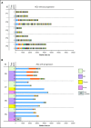
Clonality of asymptomatic HTLV-1 carriers over time. Each colored bar segment represents a unique clone, and the segment width represents the clone size. (A) Clonality among ACs who remained ACs over time. These samples exhibited polyclonal patterns (uniform distribution) over all analyzed time points. (B) Clonality among ACs who progressed to different ATL subtypes over time. These samples largely exhibited expanded oligoclonal or monoclonal patterns.
Figure 2.
Figure 2.
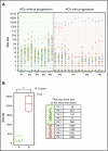
Clone size distributions among ACs. Clones are displayed in descending order based on size. Three main patterns were observed across the samples: polyclonal, oligoclonal, and monoclonal. (A) Left-most section of the graph: ACs whose clinical status did not progress. None of the samples with polyclonal patterns showed progression over time. Right-most section of the graph: ACs whose clinical status progressed. All ACs with atypical oligoclonal or monoclonal patterns showed progression. (B) Comparison of clone sizes between ACs with and without progression. Group A, ACs without progression; Group B, ACs with progression. Clone sizes of the first top clones among ACs who remained ACs vs those who developed ATL. The intergroup difference regarding the size of the first top clone was significant (Student t test, P = .0001).
Figure 3.
Figure 3.
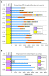
Clonality among patients with indolent-type ATL. Each colored bar segment represents a unique clone, and the segment width indicates the clone size. (A) Longitudinal analysis of clonality among patients with indolent types of ATL (SM and chronic ATL) who exhibited oligoclonal or monoclonal patterns. (B) Clonality among individuals with SM and chronic ATL who progressed to acute ATL over time. These samples largely exhibited monoclonal patterns.
Figure 4.
Figure 4.
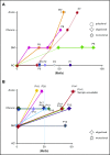
Schematic presentation of clinical courses and clonalities. (A-B) Schematic descriptions of the clinical courses of 9 asymptomatic HTLV-1 carriers and 11 patients with indolent-subtype ATL. The clinical status is shown on the vertical axis, and follow-up periods are indicated on the horizontal axis. The clonality of each patient is indicated in the figure by respective marks in the figure.

References

    1. Yoshida M, Seiki M, Yamaguchi K, Takatsuki K. Monoclonal integration of human T-cell leukemia provirus in all primary tumors of adult T-cell leukemia suggests causative role of human T-cell leukemia virus in the disease. Proc Natl Acad Sci USA. 1984;81(8):2534-2537. - PMC - PubMed
    1. Iwanaga M, Watanabe T, Yamaguchi K. Adult T-cell leukemia: a review of epidemiological evidence. Front Microbiol. 2012;3:322. - PMC - PubMed
    1. Ishitsuka K, Tamura K. Human T-cell leukaemia virus type I and adult T-cell leukaemia-lymphoma. Lancet Oncol. 2014;15(11):e517-e526. - PubMed
    1. Fujino T, Nagata Y. HTLV-I transmission from mother to child. J Reprod Immunol. 2000;47(2):197-206. - PubMed
    1. Gallo RC. The discovery of the first human retrovirus: HTLV-1 and HTLV-2. Retrovirology. 2005;2:17. - PMC - PubMed

LinkOut - more resources