. 2017 Sep 22;1(21):1815-1826.
doi: 10.1182/bloodadvances.2016002915. eCollection 2017 Sep 26.

Gene editing rescue of a novel MPL mutant associated with congenital amegakaryocytic thrombocytopenia

Affiliations

Gene editing rescue of a novel MPL mutant associated with congenital amegakaryocytic thrombocytopenia

Cédric Cleyrat et al. Blood Adv. .

Abstract

Thrombopoietin (Tpo) and its receptor (Mpl) are the principal regulators of early and late thrombopoiesis and hematopoietic stem cell maintenance. Mutations in MPL can drastically impair its function and be a contributing factor in multiple hematologic malignancies, including congenital amegakaryocytic thrombocytopenia (CAMT). CAMT is characterized by severe thrombocytopenia at birth, which progresses to bone marrow failure and pancytopenia. Here we report unique familial cases of CAMT that presented with a previously unreported MPL mutation: T814C (W272R) in the background of the activating MPL G117T (K39N or Baltimore) mutation. Confocal microscopy, proliferation and surface biotinylation assays, co-immunoprecipitation, and western blotting analysis were used to elucidate the function and trafficking of Mpl mutants. Results showed that Mpl protein bearing the W272R mutation, alone or together with the K39N mutation, lacks detectable surface expression while being strongly colocalized with the endoplasmic reticulum (ER) marker calreticulin. Both WT and K39N-mutated Mpl were found to be signaling competent, but single or double mutants bearing W272R were unresponsive to Tpo. Function of the deficient Mpl receptor could be rescued by using 2 separate approaches: (1) GRASP55 overexpression, which partially restored Tpo-induced signaling of mutant Mpl by activating an autophagy-dependent secretory pathway and thus forcing ER-trapped immature receptors to traffic to the cell surface; and (2) CRISPR-Cas9 gene editing used to repair MPL T814C mutation in transfected cell lines and primary umbilical cord blood-derived CD34+ cells. We demonstrate proof of principle for rescue of mutant Mpl function by using gene editing of primary hematopoietic stem cells, which indicates direct therapeutic applications for CAMT patients.


Conflict of interest statement

Conflict-of-interest disclosure: The authors declare no competing financial interests.

Figures

None
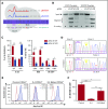
Graphical abstract
Figure 1.
Figure 1.
Novel in cis double MPL mutation is associated with familial CAMT type I. (A) Pedigree tree that illustrates the autosomal recessive transmission pattern in this family. Circles represent females and squares represent males. Open symbols indicate healthy family members, filled symbols indicate family members with CAMT type I, single horizontal line connecting 2 symbols indicates monozygotic twins, and slashes represent deceased family members. The genotypes of all family members are presented as genomic DNA sequencing chromatograms. Family members I.1, I.2, and II.1 are heterozygous for the 2 G117T mutation in exon 2 and the T814C mutation in exon 5. Family members II.2, II.3, and II.4 are homozygous for both mutations. (B) Schematic representation of the functional domains of the Tpo receptor and the location of extracellular G117T (K39N) polymorphism and T814C (W272R) mutation. C, cysteine residue; FN III, fibronectin III domain; SP, signal peptide; TM, transmembrane domain; Tyr, tyrosine residue.
Figure 2.
Figure 2.
Mpl W272R and K39N/W272R are absent from the cell surface and do not respond to ligand stimulation. (A) Western blot results for total Mpl protein and phosphorylated (p) signaling partners in cell lysates prepared from transfected Ba/F3 cells with and without Tpo stimulation (50 ng/mL, 10 minutes, 37°C). Labels at the top indicate WT or mutant MplmNG constructs for each cell line tested. (B) Representative confocal images of the live Ba/F3 cells used for western blot characterization in panel A. Closed arrowhead symbols indicate the presence of surface Mpl in cells expressing WT or K39N Mpl. Open arrowhead symbols point to the absence of a clearly defined plasma membrane outline in cells expressing W272R or K39N/W272R mutant Mpl. (C) Image analysis of human UT-7 cells co-expressing MplmNG WT or mutant proteins and the ER-resident protein calreticulin (CRT) fused to TagRFP-T (CRTTagRFP-T). Co-localization of both fluorescent markers was assessed by using Pearson’s analysis of dual-channel confocal images from at least 20 cells for each condition. Means ± standard error of the mean are shown, and pairwise statistical analyses using unpaired Student t test are represented by horizontal bars. Representative images of each cell population are shown at the bottom of the panel. (D) Co-immunoprecipitation (IP) of mutant Mpl proteins with ER-resident proteins CRT and calnexin (CANX) in stably transfected UT-7 cells. Upper bands in the WT and K39N (KN) lanes represent fully glycosylated receptors, indicative of maturation in the Golgi. Scale bars = 5 µm. *P < .05; ***P < .0001. n.s., not significant.
Figure 3.
Figure 3.
Gene editing or autophagic delivery of mutant Mpl to the cell surface rescue receptor function in vitro. (A) Transient overexpression of GRASP55 tagged with a V5 epitope results in accumulation of the lower-molecular-weight core-glycosylated form of Mpl regardless of WT or mutant status. Receptors are shown to be signaling competent on the basis of phosphorylation of key signaling proteins in the Jak/STAT and PI3K pathways in response to Tpo. (B-C) XTT-II proliferation assays performed on UT-7 or Ba/F3 cell lines expressing WT or mutant MplmNG and selected for growth in the presence of Tpo (panel C, solid lines) or eltrombopag (Elt) (panel C, dotted lines). CRISPR-Cas9–edited cells that were reverse-engineered to restore the WT sequences in MPL exon 5 from the mutated W272R sequence (labeled Mpl W272R>WT) are represented by blue open circles. UT-7 cells were edited by using the D10A Cas9 mutant and 2 single gRNAs in a double nickase approach. A classical WT Cas9 approach (ie, coupled to a unique single gRNA) was used to edit Ba/F3 cells. **P < .005; ***P < .0001.
Figure 4.
Figure 4.
Gene editing in K562 cells and primary CD34+ cells. (A) Schematic of sequence-specific gRNA#1WT and gRNA#1WR and their target genomic MPL sequence representing the protospacer-adjacent motif (PAM), double-strand break site (yellow arrowheads), and the W272R single point mutation site (T814C). DNA codons are underlined, and the repair template (RT) used to convert the W272R mutation to the WT sequence (W272R>WT) is also represented. (B) Example of in vitro digestion assay with gRNA#1WT or gRNA#1WR in the presence of their match or mismatch target sequences. Quantification of cutting efficiency was performed by using densitometry analysis. (C) Left panel shows quantification of in vitro cutting capabilities of gRNA#1WT and gRNA#1WR. Right panel shows quantification of the percentage of indel formation obtained with gRNA#1WT and gRNA#1WR when delivered as plasmid DNA or RNP complexes in K562 or CB CD34+ cells. (D) Control, unedited, and edited CD34+ cells isolated from patient II.4 were sequenced at day 5 after editing. G117T represents the K39N mutation and T814C represents the W272R mutation. Dotted magenta rectangles highlight the presence of additional overlapping sequences in edited cells for the T814C locus, indicating an off-target effect. (E) Flow cytometry analysis of anti-Mpl (CD110)-AlexaFluor-647 binding on control CD34+ cells, unedited patient II.4 CD34+ cells, or edited II.4 CD34+ cells at day 5 after editing. (F) In vitro megakaryocytic colony formation assay conducted in the presence of Tpo with the same cell samples used in panel E. *P < .05; **P < .005; ***P < .0001. CFU, colony-forming unit; DSB, double-strand break.
Figure 5.
Figure 5.
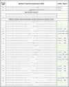
Next-generation sequencing of MPL exon 5 PCR amplicons after gene editing. Amplicons generated from gDNA obtained from patient II.4 edited CD34+ cells were subjected to next-generation sequencing. Sequences, other than properly HDR edited or unedited, that can potentially yield functional Mpl proteins are indicated in green.
Figure 6.
Figure 6.
Functional rescue strategies for Mpl mutants. Schematic summary of the 2 rescue approaches used to restore Mpl function: (1) overexpression of GRASP55 to force immature Mpl receptor expression at the cell surface using unconventional autophagy-dependent secretion and (2) CRISPR-Cas9 gene editing to convert mutated Mpl DNA sequence to WT sequence. sgRNA, single-guide RNA.

References

    1. Ihara K, Ishii E, Eguchi M, et al. Identification of mutations in the c-mpl gene in congenital amegakaryocytic thrombocytopenia. Proc Natl Acad Sci USA. 1999;96(6):3132-3136. - PMC - PubMed
    1. Bennett M, Stroncek DF. Recent advances in the bcr-abl negative chronic myeloproliferative diseases. J Transl Med. 2006;4:41. - PMC - PubMed
    1. He X, Chen Z, Jiang Y, Qiu X, Zhao X. Different mutations of the human c-mpl gene indicate distinct haematopoietic diseases. J Hematol Oncol. 2013;6:11. - PMC - PubMed
    1. Tijssen MR, di Summa F, van den Oudenrijn S, et al. Functional analysis of single amino-acid mutations in the thrombopoietin-receptor Mpl underlying congenital amegakaryocytic thrombocytopenia. Br J Haematol. 2008;141(6):808-813. - PubMed
    1. Varghese LN, Zhang JG, Young SN, et al. Functional characterization of c-Mpl ectodomain mutations that underlie congenital amegakaryocytic thrombocytopenia. Growth Factors. 2014;32(1):18-26. - PubMed