. 2017 Oct 10;1(22):1923-1933.
doi: 10.1182/bloodadvances.2017007435.

Pak2 regulates myeloid-derived suppressor cell development in mice

Affiliations

Pak2 regulates myeloid-derived suppressor cell development in mice

Yi Zeng et al. Blood Adv. .

Abstract

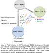
Myeloid-derived suppressor cells (MDSCs) are CD11b+Gr1+ cells that induce T-cell hyporesponsiveness, thus impairing antitumor immunity. We have previously reported that disruption of Pak2, a member of the p21-activated kinases (Paks), in hematopoietic stem/progenitor cells (HSPCs) induces myeloid lineage skewing and expansion of CD11bhighGr1high cells in mice. In this study, we confirmed that Pak2-KO CD11bhighGr1high cells suppressed T-cell proliferation, consistent with an MDSC phenotype. Loss of Pak2 function in HSPCs led to (1) increased hematopoietic progenitor cell sensitivity to granulocyte-macrophage colony-stimulating factor (GM-CSF) signaling, (2) increased MDSC proliferation, (3) decreased MDSC sensitivity to both intrinsic and Fas-Fas ligand-mediated apoptosis, and (4) promotion of MDSCs by Pak2-deficient CD4+ T cells that produced more interferon γ, tumor necrosis factor α, and GM-CSF. Pak2 disruption activated STAT5 while downregulating the expression of IRF8, a well-described myeloid transcription factor. Together, our data reveal a previously unrecognized role of Pak2 in regulating MDSC development via both cell-intrinsic and extrinsic mechanisms. Our findings have potential translational implications, as the efficacy of targeting Paks in cancer therapeutics may be undermined by tumor escape from immune control and/or acceleration of tumorigenesis through MDSC expansion.


Conflict of interest statement

Conflict-of-interest disclosure: The authors declare no competing financial interests.

Figures
None
Graphical abstract
Figure 1.
Figure 1.
Genetic disruption of Pak2 in HSPCs induces MDSC expansion in the spleen. CD45.1+ naive BoyJ mice were noncompetitively transplanted with CD45.2+Pak2flox/floxMx1Cre+ (Pak2-KO) or CD45.2+Pak2flox/floxMx1Cre (WT) BM cells and treated with polyIC. (A) Gr1highLy6G+ cells that were isolated from spleens suppressed T-cell proliferation. (B) The numbers of phenotypic granulocytic (CD11b+Ly6G+Ly6Clow) and monocytic (CD11b+Ly6Glow/−Ly6Chi) MDSCs are shown per spleen. (C-D) WT (C) and Pak2-KO (D) splenic CD45.2+Gr1highLy6G+ cells were stained with Giemsa. Inserts show morphology of cells at a high magnification. All scale bars represent 20 mm. Representative data from at least 3 experiments with 3 to 9 mice per genotype are shown.
Figure 2.
Figure 2.
Pak2-deficient MDSCs are more suppressive than MDSCs from tumor-bearing mice. Mice reconstituted with WT and Pak2-KO BM were inoculated with LLC cells or PBS control subcutaneously. (A) The numbers of CD45.2+CD11b+Gr1high (representing PMNs and MDSCs) and CD45.2+CD11b+Gr1low cells (representing monocytes) are shown per spleen. (B) Effects of splenic Gr1highLy6G+ cells on T-cell proliferation. Representative data from 2 experiments with 5 to 9 mice per genotype are shown. *P < .05; **P < .01; ***P < .001; ****P < .0001; ns, not significant.
Figure 3.
Figure 3.
Pak2-KO HPCs display increased sensitivity to GM-CSF signaling. (A) MDSCs were generated from Pak2-KO or WT BM cells in the presence of GM-CSF, IL-6, and G-CSF. (B) Colony formations of sorted CD45.2+ BM cells cultured in methylcellulose with GM-CSF. The numbers of colonies (colony-forming units in response to GM-CSF [CFU-GM]) per femur are shown. (C) GM-CSF receptor α (Csf2ra) and β (Csf2rb) chain gene expression was measured in progenies collected from the GM-CSF colony assay shown in Figure 3B. Pak2-KO or WT BM c-kit+ cells were cultured with GM-CSF for 7 days. (D-E) Cells were stained for CD11b and Gr1 (D) and cocultured with T cells to test their suppressive function (E). The suppressive function on CD8+ T-cell proliferation is shown. Representative data from at least 3 experiments with 3 to 6 mice per genotype are shown. **P < .01; ***P < .001.
Figure 4.
Figure 4.
Pak2-KO spleen MDSCs are more proliferative than WT cells Freshly isolated splenocytes from mice reconstituted with Pak2-KO or WT BM were stained for CD11b, Gr1, and intracellular Ki-67. (A) MFI of Ki-67 in gated CD45.2+CD11bhighGr1high and CD11b+Gr1low populations is shown. (B) Representative flow histograms are shown. Representative data from 2 experiments with 3 to 6 mice per genotype are shown. ***P < .001.
Figure 5.
Figure 5.
Pak2 disruption results in decreased spontaneous and Fas-FasL–induced apoptosis in MDSCs. (A) Freshly isolated splenocytes from mice reconstituted with Pak2-KO or WT BM were cultured in the presence or absence of FasL for 2 hours prior to CD11b, Gr1, Annexin V, and propidium iodide (PI) staining. The percentage of Annexin V+PI+ cells in the gated CD45.2+CD11bhighGr1high population is shown. (B) Freshly isolated splenocytes from mice reconstituted with Pak2-KO or WT BM were stained for CD45.2, Fas, CD11b, Gr1, CD3, and B220. The MFI of Fas in gated CD45.2+ populations is shown. (C) Bcl2, (D) Mcl1, (E) Bcl-xL, (F) Bak1, and (G) Bax expression in Pak2-KO MDSCs or WT PMNs (progenies from the GM-CSF colony assays shown in Figure 3B) was determined by quantitative real-time PCR. Representative data from 3 or 4 experiments with 3 to 10 mice per genotype are shown. *P < .05; **P < .01; ***P < .001; ****P < .0001.
Figure 6.
Figure 6.
Loss of Pak2 activates STAT5 and downregulates IRF8 expression in MDSCs. (A) STAT5a gene expression and (B) STAT5 protein phosphorylation in Pak2-KO MDSCs or WT PMNs (progenies from the GM-CSF colony assays shown in Figure 3B) were determined by quantitative real-time PCR and flow cytometry, respectively. (C-D) IRF8 gene expression in Pak2-KO MDSCs or WT PMNs (progenies from the GM-CSF colony assays shown in Figure 3B) (C) and Pak2-KO or WT Gr1highLy6G+ cells (MDSCs and PMNs, respectively) (D) isolated from spleen are shown. Representative data from 2 or 3 experiments with 3 to 6 mice per genotype are shown. *P < .05; ****P < .0001.
Figure 7.
Figure 7.
Pak2-deficient T cells induce MDSC generation in vitro. Purified splenic CD4+ T cells from mice reconstituted with Pak2-KO or WT BM were stimulated with CD3/CD28 beads for 3 days before supernatant was collected. (A-C) Amounts of GM-CSF (A), TNF-α (B), and IFN-γ (C) in the supernatant were determined by enzyme-linked immunosorbent assay. Naive C57BL/6 mice BM cells were cultured in the presence of supernatant from Pak2-KO or WT splenic CD4+ T cells for 5 days, collected, and then cocultured with purified splenic T cells from naive C57BL/6 mice to measure their suppressive function on T-cell proliferation. (D-E) The percentage of the CD11bhighGr1high population in panel D and the percentage suppression of T-cell proliferation by BM cells cultured in CD4+ T-cell supernatant (E) are shown. Representative data from 2 or 3 experiments with 3 to 6 mice per genotype are shown. *P < .05; **P < .01; ***P < .001; ***P < .0001.

References

    1. Gabrilovich DI, Nagaraj S. Myeloid-derived suppressor cells as regulators of the immune system. Nat Rev Immunol. 2009;9(3):162-174. - PMC - PubMed
    1. Nagaraj S, Collazo M, Corzo CA, et al. . Regulatory myeloid suppressor cells in health and disease. Cancer Res. 2009;69(19):7503-7506. - PMC - PubMed
    1. Gabrilovich DI, Ostrand-Rosenberg S, Bronte V. Coordinated regulation of myeloid cells by tumours. Nat Rev Immunol. 2012;12(4):253-268. - PMC - PubMed
    1. Youn JI, Collazo M, Shalova IN, Biswas SK, Gabrilovich DI. Characterization of the nature of granulocytic myeloid-derived suppressor cells in tumor-bearing mice. J Leukoc Biol. 2012;91(1):167-181. - PMC - PubMed
    1. Youn JI, Nagaraj S, Collazo M, Gabrilovich DI. Subsets of myeloid-derived suppressor cells in tumor-bearing mice. J Immunol. 2008;181(8):5791-5802. - PMC - PubMed