. 2018 Jan 18;378(3):250-261.
doi: 10.1056/NEJMoa1709449. Epub 2018 Jan 3.

Somatic Activating KRAS Mutations in Arteriovenous Malformations of the Brain

Affiliations

Somatic Activating KRAS Mutations in Arteriovenous Malformations of the Brain

Sergey I Nikolaev et al. N Engl J Med. .

Abstract

Background: Sporadic arteriovenous malformations of the brain, which are morphologically abnormal connections between arteries and veins in the brain vasculature, are a leading cause of hemorrhagic stroke in young adults and children. The genetic cause of this rare focal disorder is unknown.

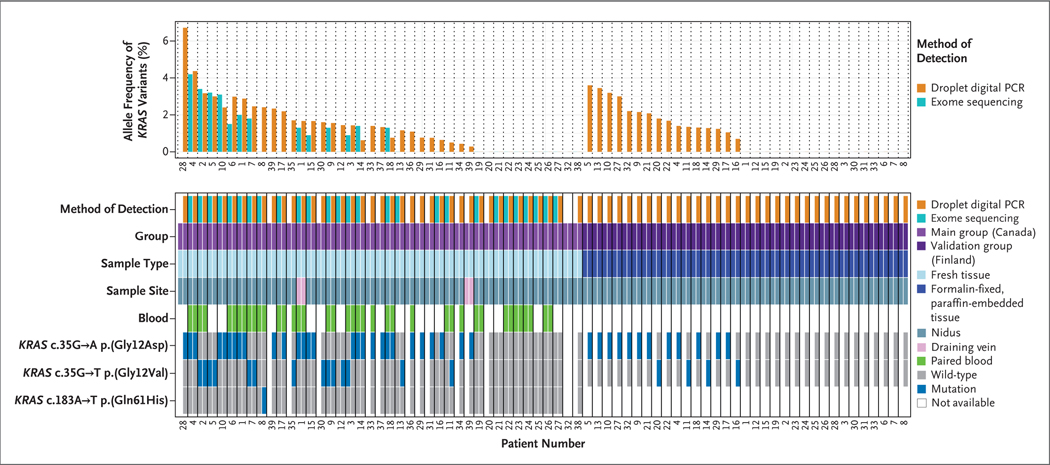
Methods: We analyzed tissue and blood samples from patients with arteriovenous malformations of the brain to detect somatic mutations. We performed exome DNA sequencing of tissue samples of arteriovenous malformations of the brain from 26 patients in the main study group and of paired blood samples from 17 of those patients. To confirm our findings, we performed droplet digital polymerase-chain-reaction (PCR) analysis of tissue samples from 39 patients in the main study group (21 with matching blood samples) and from 33 patients in an independent validation group. We interrogated the downstream signaling pathways, changes in gene expression, and cellular phenotype that were induced by activating KRAS mutations, which we had discovered in tissue samples.

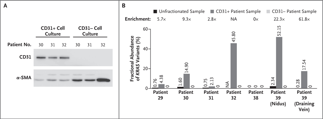
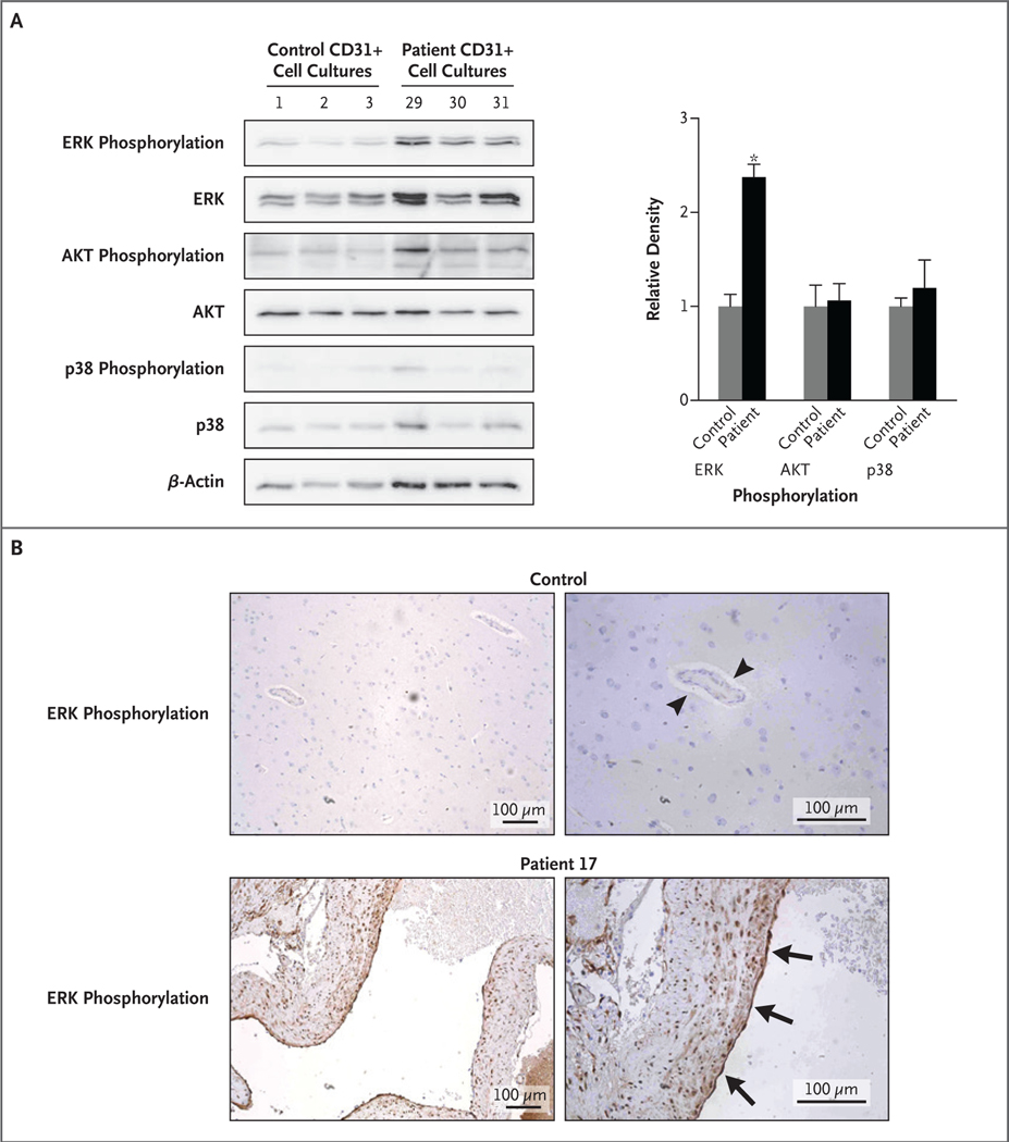
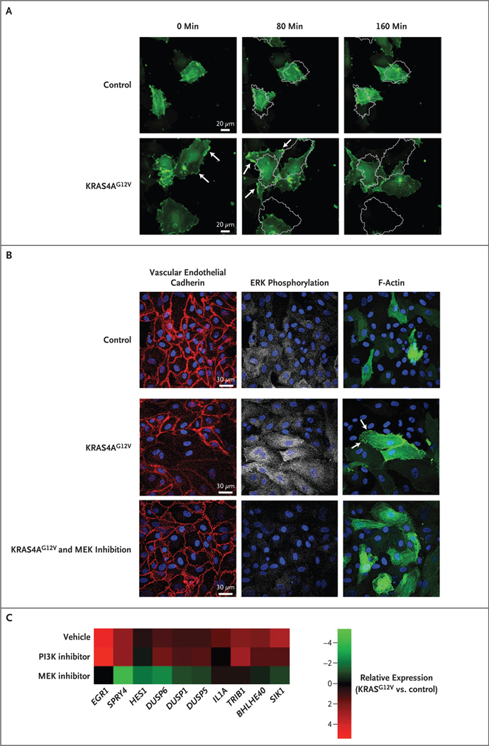
Results: We detected somatic activating KRAS mutations in tissue samples from 45 of the 72 patients and in none of the 21 paired blood samples. In endothelial cell-enriched cultures derived from arteriovenous malformations of the brain, we detected KRAS mutations and observed that expression of mutant KRAS (KRASG12V) in endothelial cells in vitro induced increased ERK (extracellular signal-regulated kinase) activity, increased expression of genes related to angiogenesis and Notch signaling, and enhanced migratory behavior. These processes were reversed by inhibition of MAPK (mitogen-activated protein kinase)-ERK signaling.

Conclusions: We identified activating KRAS mutations in the majority of tissue samples of arteriovenous malformations of the brain that we analyzed. We propose that these malformations develop as a result of KRAS-induced activation of the MAPK-ERK signaling pathway in brain endothelial cells. (Funded by the Swiss Cancer League and others.).


Figures

Figure 1.
Figure 1.. Detection of KRAS Mutations in Samples Obtained from Patients.
The top chart shows the allele frequency of KRAS variants, determined by either the percentage of sequence reads that contained variants on whole-exome sequencing or the fractional abundance of variants on digital droplet polymerase-chain-reaction (PCR) analysis, in tissue samples of arteriovenous malformations of the brain. The samples are shown according to patient number in order of highest to lowest frequency, first in the main study group, which included 39 patients from Canada (29 [74%] with KRAS mutations), and then in the independent validation group, which included 33 patients from Finland (16 [48%] with KRAS mutations). The bottom chart shows details about the samples, including the sample type (fresh-frozen or formalin-fixed, paraffin-embedded tissue), sample site (nidus or draining vein), the presence or absence of a paired blood sample, and the specific activating mutation detected. No KRAS mutations were detected in paired blood samples. The CD31+ cells from Patient 32 in the main study group were positive for a c.35G→A KRAS mutation (data not shown in this figure), but there was insufficient tissue remaining for whole tissue analysis. Two patients from the main study group (Patients 1 and 39) had KRAS mutations in the draining-vein sample that matched those observed in the nidus sample.
Figure 2.
Figure 2.. Detection of KRAS Mutations in Endothelial-Cell–Enriched and Endothelial-Cell–Depleted Cultures.
Panel A shows the expression of CD31 and alpha smooth-muscle actin (α-SMA) in endothelial-cell–enriched (CD31+) and endothelial-cell–depleted (CD31−) fractions of cell cultures derived from tissue samples of arteriovenous malformations of the brain that were obtained from patients. The CD31+ fractions (isolated with anti-CD31 magnetic beads) were composed of CD31+ cells and some α-SMA+ cells, whereas the CD31− fractions were devoid of CD31+ cells but contained α-SMA+ cells. Panel B shows the fractional abundance of KRAS variants in CD31+ and CD31− cell cultures derived from tissue samples of arteriovenous malformations of the brain and in the whole tissue sample before fractionation. Enrichment is the factor change of the fractional abundance in the CD31+ cells as compared with the whole unfractionated sample. The samples are from patients in the main study group. For Patient 32, all available tissue was used for cell culture and no tissue was left for digital droplet polymerase-chain-reaction analysis. NA denotes not available.
Figure 3.
Figure 3.. Detection of ERK Phosphorylation in Endothelial-Cell–Enriched Cell Cultures and Tissue Samples.
In Panel A, endothelial-cell–enriched (CD31+) cell cultures derived from tissue samples of arteriovenous malformations of the brain with KRAS mutations (from Patients 29, 30, and 31 in the main study group) show increased phosphorylation of ERK1/2 (extracellular signal-regulated kinase 1 or 2) but not of p38 or AKT, whereas three control CD31+ cell cultures derived from normal brain vasculature do not show increased phosphorylation of ERK1/2. Densitometry is shown on the right, normalized to beta-actin. The asterisk indicates a P value of 0.002. In Panel B, immunohistochemical staining of a tissue sample of an arteriovenous malformation of the brain with a KRAS mutation (from Patient 17 in the main study group) shows strong staining for ERK phosphorylation in endothelial cells lining the vascular lumen (arrows) and in vascular smooth-muscle cells in the vessel wall, whereas normal brain parenchymal vessels show little or no staining for ERK phosphorylation in endothelial cells (arrowheads).
Figure 4.
Figure 4.. Phenotype of Endothelial Cells Expressing Active KRAS.
Panel A shows time-lapse confocal images of human umbilical-vein endothelial cells (HUVECs) with KRAS4AG12V expression and control cells. In the absence of exogenous angiogenic factors or a migratory cue (e.g., scratch wound), KRAS4AG12V expression altered the actin dynamics (assessed by cotransfected LifeAct-GFP), including a reduction in the number of filopodia and an increase in lamellipodia formation (lamellipodia are indicated by arrows). Actin turnover was more rapid, and cells had increased motility; the images obtained at 80 minutes and 160 minutes show a gray outline of the position of the cells at 0 minutes. Panel B shows staining for vascular endothelial cadherin, ERK phosphorylation, and F-actin (assessed by cotransfected LifeAct-GFP) in HUVECs with KRAS4AG12V expression, HUVECs with KRAS4AG12V expression and MAPK–ERK inhibition with a MEK inhibitor for 16 hours, and control cells. KRAS4AG12V activated the MAPK–ERK pathway and promoted disassembly of adherens junctions. MAPK–ERK inhibition re-established the formation of adherens junctions and normalized actin localization (arrows) and cell shape. Panel C is a heat map showing the expression of vascular endothelial growth factor (VEGF)–regulated genes in HUVECs expressing KRAS4AG12V, treated with vehicle, MEK inhibitor, or phosphoinositide 3 kinase (PI3K) inhibitor for 6 hours in serum-starvation media. Data were compared with control cells treated with vehicle.

Comment in

References

    1. Brown RD Jr, Wiebers DO, Torner JC, O’Fallon WM. Incidence and prevalence of intracranial vascular malformations in Olmsted County, Minnesota, 1965 to 1992. Neurology 1996;46: 949–52. - PubMed
    1. Berman MF, Sciacca RR, Pile-Spellman J, et al. The epidemiology of brain arteriovenous malformations. Neurosurgery 2000; 47:389–97. - PubMed
    1. Al-Shahi R, Warlow C. A systematic review of the frequency and prognosis of arteriovenous malformations of the brain in adults. Brain 2001; 124: 1900–26. - PubMed
    1. Al-Shahi R, Fang JS, Lewis SC, Warlow CP. Prevalence of adults with brain arteriovenous malformations: a community based study in Scotland using capturere-capture analysis. J Neurol Neurosurg Psychiatry 2002; 73:547–51. - PMC - PubMed
    1. McAllister KA, Grogg KM, Johnson DW, et al. Endoglin, a TGF-beta binding protein of endothelial cells, is the gene for hereditary haemorrhagic telangiectasia type 1. Nat Genet 1994; 8: 345–51. - PubMed

Publication types

MeSH terms

Substances