. 2018 Jan 2;22(1):242-254.
doi: 10.1016/j.celrep.2017.12.007.

Hair Follicle Development in Mouse Pluripotent Stem Cell-Derived Skin Organoids

Affiliations

Hair Follicle Development in Mouse Pluripotent Stem Cell-Derived Skin Organoids

Jiyoon Lee et al. Cell Rep. .

Abstract

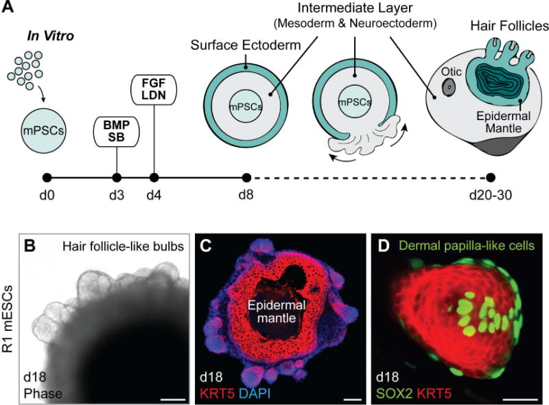
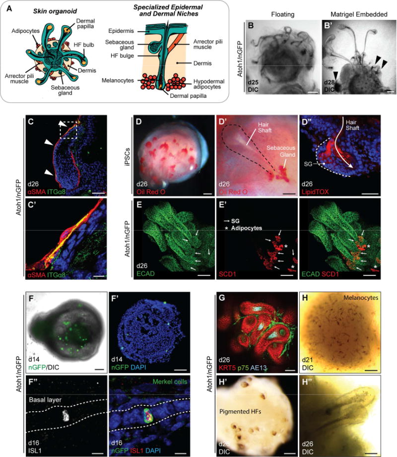
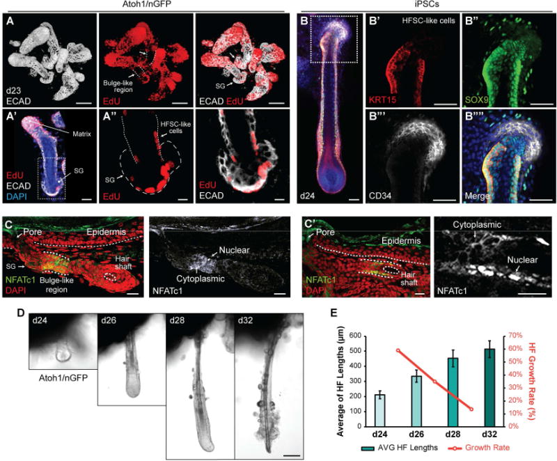
The mammalian hair follicle arises during embryonic development from coordinated interactions between the epidermis and dermis. It is currently unclear how to recapitulate hair follicle induction in pluripotent stem cell cultures for use in basic research studies or in vitro drug testing. To date, generation of hair follicles in vitro has only been possible using primary cells isolated from embryonic skin, cultured alone or in a co-culture with stem cell-derived cells, combined with in vivo transplantation. Here, we describe the derivation of skin organoids, constituting epidermal and dermal layers, from a homogeneous population of mouse pluripotent stem cells in a 3D culture. We show that skin organoids spontaneously produce de novo hair follicles in a process that mimics normal embryonic hair folliculogenesis. This in vitro model of skin development will be useful for studying mechanisms of hair follicle induction, evaluating hair growth or inhibitory drugs, and modeling skin diseases.

Keywords: 3D culture; dermis; epidermis; hair follicle; organoids; pluripotent stem cells; skin; skin appendages.


Conflict of interest statement

Declaration of Interests

J.L. and K.R.K, with the Indiana University Research and Technology Corporation, have applied for a patent related to the cell culture method described in this manuscript.

Figures

Figure 1
Figure 1. Spontaneous HF bulb development in R1 mESC aggregates. See also Figure S1
A, Schematic overview of mouse skin organoid culture. On day 0 of differentiation, dissociated mPSCs were cultured as an aggregate. On day 3, the aggregate was treated with SB and BMP to induce surface ectoderm, followed by day 4 treatment with FGF and LDN to induce placodal epithelium. By day 8, the aggregate comprises an undifferentiated PSC core, intermediate layer containing mesoderm and neuroectoderm cells, and outermost layer of surface ectoderm. From day 8, the aggregate is in a maturation medium, and undergoes an inside-out transformation as the intermediate layer erupts out to cover the surface ectoderm. B, Representative phase contrast image. On day 18, bulb-like HFs protrude from the surface of an R1 mESC aggregate. C, D, Representative IHC images. HF-like bulbs with KRT5+ epithelium in a skin organoid cryosection on day 18 (C) and a protruding KRT5+ HF-like bulb structure with SOX2+ DP-like cells (D). Scale bars, 100 µm (B, C), 50 µm (D).
Figure 2
Figure 2. Comparison of R1 and Atoh1/nGFP mESC developmental morphologies and HF formation. See also Figure S2 and Table S1
A, B, Comparison of SOX2+ neuroectoderm-like compartment and KRT5+ epithelium formation between R1 and Atoh1/nGFP mESC aggregates during developmental stages, specifically on day 14. NE; Neuroectoderm, OtV; Otic Vesicle. C, Representative DIC images of day 18 Atoh1/nGFP aggregates of each morphology category, based on the degree of the exterior tissues present outside the epithelium: uncovered, partially covered, and fully covered. D, Representative DIC images of day 28 Atoh1/nGFP aggregates of each HF-production category, based on the number of HFs produced: 1–5 HFs (*), 6–15 HFs (**), and >15 HFs (***). Arrowheads indicate HFs. E, The central pie graph represents percentages of Atoh1/nGFP aggregates in three different morphologies (Uncovered; 29/193 aggregates, Partially Covered; 117/193 aggregates, and Fully Covered; 47/193 aggregates), categorized on day 18 of each experiment. The branched pie graphs represent percentages of aggregates categorized based on the production of HFs per morphology group, on days 22–31 (Uncovered: 0, 4/29; *, 5/29; **, 4/29; ***, 16/29; Partially Covered: 0, 12/117; *, 24/117; **, 30/117; ***, 51/117; and Fully Covered: 0, 28/47; *, 19/47; **, 0/47; ***, 0/47 aggregates). Scale bars, 200 µm (C, D), 100 µm (A, B).
Figure 3
Figure 3. Self-assembly of skin layers in skin organoids. See also Figure S3
A, Schematic overview of skin developmental stages in vitro. KRT5+ basal keratinocytes are present on day 10, and KRT10+ spinous-like epidermal cells are present by day 14. During days 18–30, a Loricrin+ granular layer and a FLG+ cornified layer form. Developing HFs are visible by day 18. Dermis markers are observed on day 12 of differentiation and adipocyte-rich hypodermis forms by day 30. BE′, Representative IHC images of self-assembled epidermis during days 10–25: (B) KRT5+ basal layer, (C) KRT10+ spinous layer, (D, D) Loricrin+ granular layer, and (E, E) FLG+ cornified layer. (D, E, E) TUJ1+ neuroectoderm cells are denoted NE. FG′, Representative IHC images of dermis developed on days 12–18: (F) DLK1+ and (G) CD34+ dermal cells are formed surrounding the epidermis, and (F) COL3α1 and (G) COL4α1 are localized at the epidermal-dermal interface. HI′, Representative hypodermis images. Adipocytes developed in the hypodermal layer are visualized by (H) Oil Red O and (I, I) LipidTOX stainings. Arrowheads (H) indicate two different protruding HFs. Dash-lined boxes (F, G, I) indicate the area of magnification. Scale bars: 100 µm (BD, EI), 10 µm (D).
Figure 4
Figure 4. Stages of HF induction recapitulated in Atoh1/nGFP mESCs and miPSCs. See also Figure S4
A, Illustration of the first four of eight stages of native HF development: hair germ, peg, and bulb formation. B, Representative IHC images of EDAR+ LHX2+ developing hair germs on days 14, 16, and 18. C, C′, Ki67+ cells in the hair germ and nascent HF matrix epithelium on day 26. Day 26 organoid contains HFs at different developmental stages (hair germs, pegs, and bulb-like hair pegs) in a single organoid unit. DD″, Day 18 skin organoid contains HFs with SOX2+ DP (D, *) and SOX2 germ stage HFs (D, arrowheads). Sparse SOX2+ interfollicular epithelial cells represent Merkel cells (D, circles). See also Figure 5F–F″. EE″, On day 28, skin organoids have HFs containing or lacking SOX2+ DP; HFs with SOX2 DP suggest a zigzag HF identity (E), while HFs with SOX2+ DP suggest guard, awl, or auchene HF identity (E). FI′, Representative images of developing HFs showing (F) αSMA+ dermal sheath cells, (G) KRT5+ p63+ outer root sheath and p63+ matrix cells. (H) Representative DIC image of an iPSC-derived HF on day 30. (I, I) GATA3+ inner root sheaths comprised of Huxley’s and cuticle layers with Henle’s layer, distinguishable by morphology. AE13+ cells were located in the hair cortex region of organoid HF shafts. JJ″, TEM images of the Atoh1/nGFP ESC-derived skin organoid HF on day 26. (J) Cross-section of the hair shaft showing HF lineages. From the outermost layer to the center of the HF: DS, dermal sheath; ORS, outer root sheath; Cp, companion layer; He, Henle’s layer; Hu, Huxley’s layer; Ci, inner root sheath cuticle; Ch, hair shaft cuticle; Co, cortex; Me, medulla. (J) High magnification view of the cortex and medulla. Dash-lined boxes (E, J) indicate the area of magnification. Scale bars, 100 µm (B, C, DE), 50 µm (C, FI′, J), 10 µm (J), 5 µm (J″).
Figure 5
Figure 5. Specialized cutaneous cell types in skin organoids. See also Figure S4
A, Schematic of specialized epidermal and dermal niches within skin organoids. (left) Late stage (>d20) skin organoids consist of epidermis, dermis, and HFs. (right) A close view of a single HF with sebaceous gland, arrector pili muscle, HF bulge, melanocytes in the hair shaft, and hypodermal adipocytes. B, B′, DIC images of HFs with entangled morphology in floating culture (B) and HFs with protruded morphology in Matrigel droplet (B). Arrowheads denote abnormal HFs. C, C′, αSMA+ ITGα8+ myocyte-like cells are occasionally observed in association with organoid HF dermal sheaths. Arrowheads denote αSMA and ITGα8 double-positive regions. DE′, SGs near hair shafts were labeled by Oil Red O (D, D), LipidTOX staining (D), and whole-mount staining (E, E). Arrows indicate SGs (SCD1+ ECAD+). Asterisks indicate adipocytes (SCD1+ ECAD). SG; sebaceous gland. FF″, Representative images of Merkel cells. DIC (F) and IHC (F) images on day 14 show intermittent nGFP+ cells, indicative of Merkel cells. (F) nGFP+ Merkel-like cells are also Islet1 (ISL1)-positive. G, A day 26 organoid with KRT5+ HFs, p75+ neural crest-like cells, and AE13+ HF cortex. p75 also labels dermal papilla cells. HH″, Representative DIC images of melanocyte-like cells in the epidermis on day 21 (H) and pigmented HFs on day 26 (H′, H). Dash-lined box (C) indicates the area of magnification. Scale bars: 250 µm (B), 100 µm (B, E, E′, F, F′, H, H), 50 µm (C, DD″, G, H), 10 µm (C′, F).
Figure 6
Figure 6. Development of HF bulge-like region and HF degeneration in skin organoids. See also Figure S4
AA″, Representative images of HFs on day 23, which were incubated with EdU solution for 24 hours. EdU incorporation was noted in the HF matrix (A), in the outer root sheath, and the SG (A′, A). Arrows indicate HF bulge-like region. SG; sebaceous gland, HFSC; hair follicle stem cell. BB″″, KRT15+ SOX9+ CD34 HFSC-like cells at early postnatal stage of maturity were present in the HF bulge-like region on day 24. C, C′ Representative IHC images of NFATc1 in the bulge-like region of two different Atoh1/nGFP-derived HFs. NFATc1 expression localized to the cytosol or nuclei of cells within the bulge-like region. D, E, HF morphologies were tracked during days 24–32 (D) after embedding aggregates in Matrigel on day 20 to fix aggregate position. Nine individual HFs were tracked to measure HF lengths, and HF growth rates were analyzed (E). HFs grew until day 32 while the growth rates of each interval were decreasing (D, E). HFs appeared to undergo degeneration starting on day 28 with protruding dermal papilla morphology, and eventually loosing integrity of the cells at the hair bulb region by day 32 (D). Dash-lined boxes (A′, B) indicate the area of magnification. Bars denote ± SEM. Scale bars, 100 µm (A, D), 50 µm (BB″″), 30 m (A′, C, C), 10 µm (A).

References

    1. Asakawa K, Toyoshima KE, Ishibashi N, Tobe H, Iwadate A, Kanayama T, Hasegawa T, Nakao K, Toki H, Noguchi S, Ogawa M, Sato A, Tsuji T. Hair organ regeneration via the bioengineered hair follicular unit transplantation. Sci Rep. 2012;2:424. - PMC - PubMed
    1. Chen KG, Mallon BS, McKay RDG, Robey PG. Human pluripotent stem cell culture: considerations for maintenance, expansion, and therapeutics. Cell Stem Cell. 2014;14:13–26. - PMC - PubMed
    1. Chuong CM, Cotsarelis G, Stenn K. Defining hair follicles in the age of stem cell bioengineering. J Invest Dermatol. 2007;127:2098–2100. - PMC - PubMed
    1. Coraux C, Hilmi C, Rouleau M, Spadafora A, Hinnrasky J, Ortonne JP, Dani C, Aberdam D. Reconstituted skin from murine embryonic stem cells. Curr Biol. 2003;13:849–853. - PubMed
    1. Cotsarelis G, Sun TT, Lavker RM. Label-retaining cells reside in the bulge area of pilosebaceous unit: implications for follicular stem cells, hair cycle, and skin carcinogenesis. Cell. 1990;61:1329–1337. - PubMed

Publication types