. 2018 Jan 3;10(422):eaam7577.
doi: 10.1126/scitranslmed.aam7577.

Intravenous delivery of oncolytic reovirus to brain tumor patients immunologically primes for subsequent checkpoint blockade

Affiliations

Intravenous delivery of oncolytic reovirus to brain tumor patients immunologically primes for subsequent checkpoint blockade

Adel Samson et al. Sci Transl Med. .

Abstract

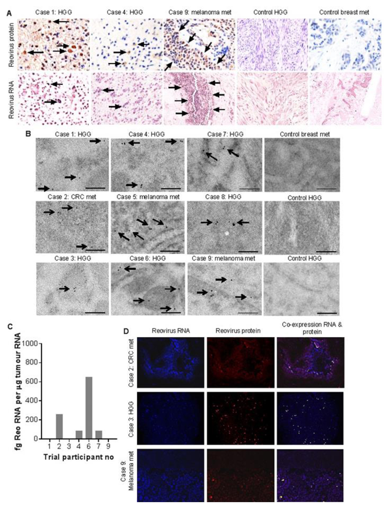
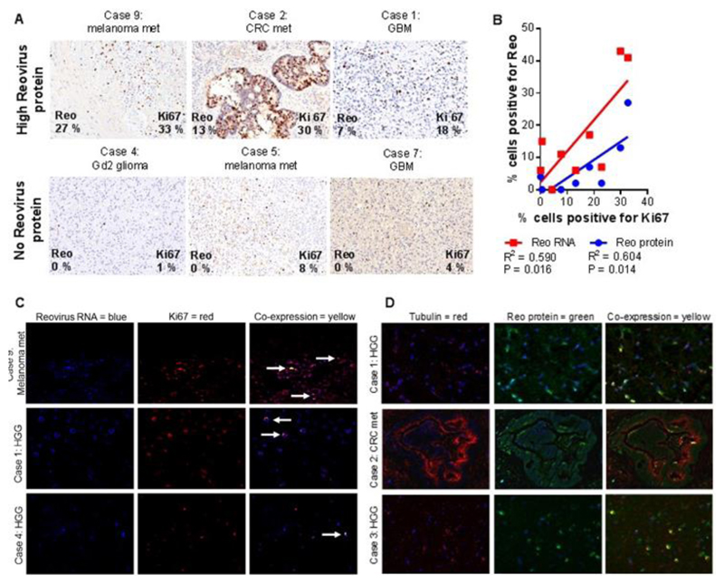
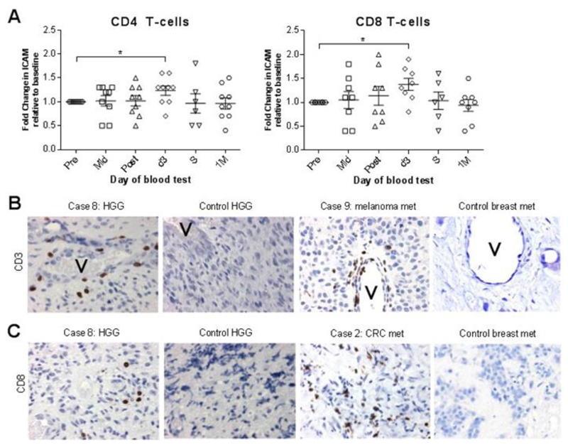
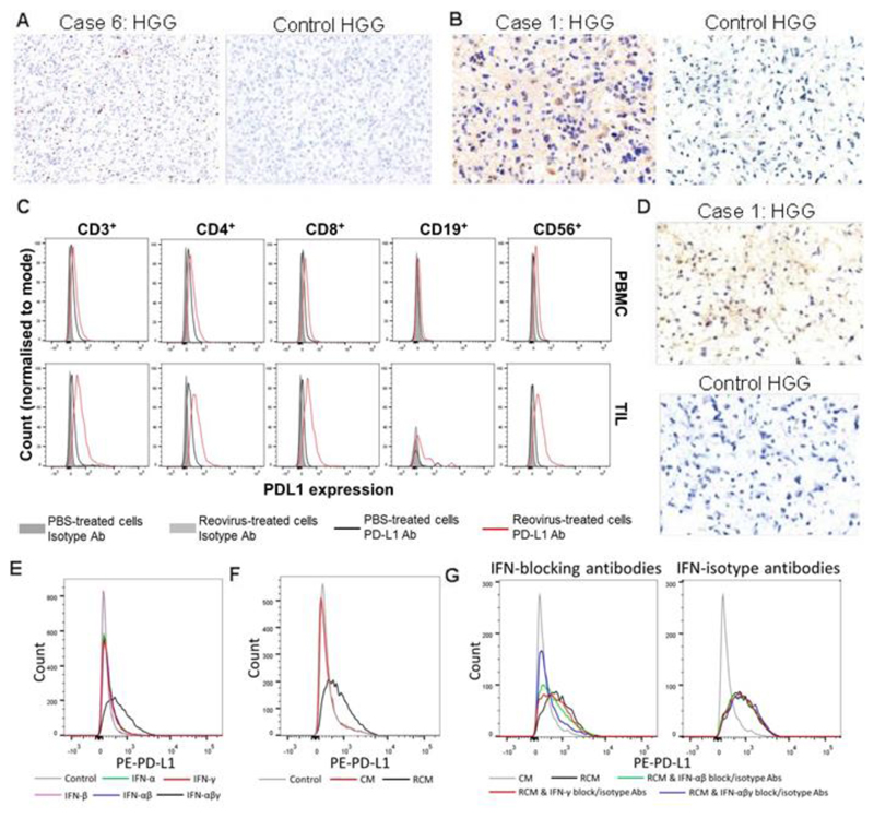
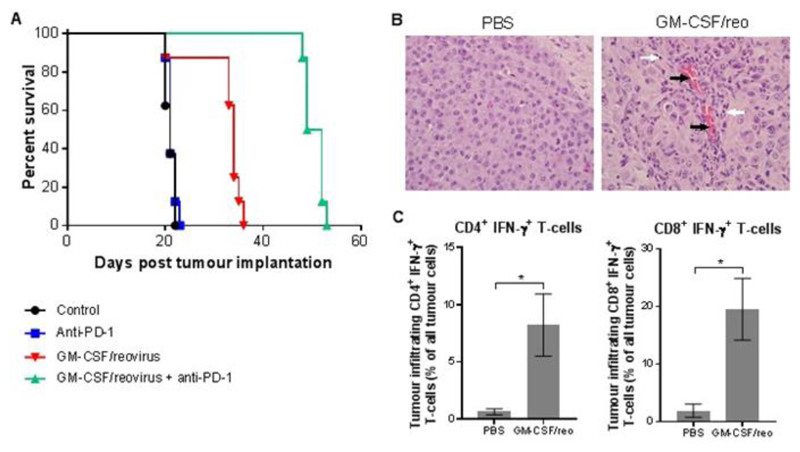
Immune checkpoint inhibitors, including those targeting programmed cell death protein 1 (PD-1), are reshaping cancer therapeutic strategies. Evidence suggests, however, that tumor response and patient survival are determined by tumor programmed death ligand 1 (PD-L1) expression. We hypothesized that preconditioning of the tumor immune microenvironment using targeted, virus-mediated interferon (IFN) stimulation would up-regulate tumor PD-L1 protein expression and increase cytotoxic T cell infiltration, improving the efficacy of subsequent checkpoint blockade. Oncolytic viruses (OVs) represent a promising form of cancer immunotherapy. For brain tumors, almost all studies to date have used direct intralesional injection of OV, because of the largely untested belief that intravenous administration will not deliver virus to this site. We show, in a window-of-opportunity clinical study, that intravenous infusion of oncolytic human Orthoreovirus (referred to herein as reovirus) leads to infection of tumor cells subsequently resected as part of standard clinical care, both in high-grade glioma and in brain metastases, and increases cytotoxic T cell tumor infiltration relative to patients not treated with virus. We further show that reovirus up-regulates IFN-regulated gene expression, as well as the PD-1/PD-L1 axis in tumors, via an IFN-mediated mechanism. Finally, we show that addition of PD-1 blockade to reovirus enhances systemic therapy in a preclinical glioma model. These results support the development of combined systemic immunovirotherapy strategies for the treatment of both primary and secondary tumors in the brain.


Conflict of interest statement

Competing interests: MC is an employee of Oncolytics Biotech Inc., Calgary, Canada, from which SG, SCS, AM, KH and HP have received research grants. MC and colleagues are inventors on several patents (including patents 6110461, 6136307, 6261555, 6344195, 6455038) held by Oncolytics Biotech Inc. that cover reovirus treatment of neoplasia. All other authors declare that they have no competing interests.

Figures

Fig. 1
Fig. 1. Intravenous delivery of reovirus to primary and secondary brain tumors.
A) Representative IHC and ISH trial and control patient tumor sections stained for reovirus σ3 protein (brown - top row) and reovirus RNA (blue - bottom row). The ‘control breast met’ is a metastasis to the brain from a breast cancer primary. Arrows point to examples of positive cells / positive areas of tissue. Scale bars = 20 μm. B) Trial and control patient tumor immunogold-TEM images for reovirus σ3 protein (arrows). Scale bar = 200 nm. C) qRT-PCR for reovirus σ3 gene, using whole tumor RNA. Data indicate fg reovirus RNA per µg of whole tumor RNA. Histogram shows the mean of triplicate samples, and error bars indicate standard deviation. D) Representative IF of trial patient tumor sections showing staining for reovirus RNA (blue), reovirus σ3 protein (red), and their co-expression (yellow). Scale bars = 80 μm.
Fig. 2
Fig. 2. Correlation of reovirus RNA / protein with proliferating tumor cells
A) Trial patient IHC tumor sections stained for Ki67 (brown) with indicated percentages of cells positive for Ki67 and reovirus σ3 protein (from table S3), showing examples of tumors with high reovirus σ3 staining (top row) and no reovirus σ3 protein staining (bottom row). Scale bars = 40 μm. B) Scatter plot and line of best fit, correlating the percentages of tumor cells positive by IHC for reovirus RNA or σ3 protein and for Ki67. C) Representative tumor sections derived from trial patient nine (high Ki67, top row), trial patient one (intermediate Ki67, middle row), and trial patient four (low Ki67, bottom row), showing IF staining for reovirus RNA (blue), Ki67 (red), or their co-expression (yellow, arrows). Scale bars = 40 μm. D) Representative trial patient tumor IF staining for tubulin (fluorescent red), reovirus σ3 protein (fluorescent green) and their co-expression (yellow). Nuclear counterstaining is blue. Top and bottom row scale bars = 40 μm. Middle row scale bar = 80 μm.
Fig. 3
Fig. 3. Tumor immune cell infiltration
A) Fold change in cell-surface ICAM expression on CD4 and CD8 T cells from trial patients’ peripheral blood. B) Trial and control patient IHC tumor sections stained for CD3 (brown). Scale bars = 20 μm. ‘V’ indicates blood vessel. C) Trial and control patient IHC tumor sections stained for CD8 (brown). Scale bars = 20 μm.
Fig. 4
Fig. 4. Expression of cleaved caspase 3, PD-L1, and PD-1 in high grade gliomas after reovirus treatment
A) Representative trial and control patient HGG sections stained for cleaved caspase 3 (brown) by IHC. Scale bars = 60 μm. B) Representative trial and control patient HGG sections stained by IHC for PD-L1 (brown). Scale bars = 30 μm. C) Representative (one of three samples tested) flow cytometry for PD-L1 on GBM TILs (bottom row) or PBMCs (top row) derived from the same patient, after stimulation for 48 hours using 1 PFU/cell reovirus. D) Representative trial and control patient HGG sections stained by IHC for PD-1 (brown). Scale bars = 30 μm. E) Representative flow cytometry for PD-L1 on GBM1 cells after stimulation with combinations of purified IFN-α/-β/-γ for 24 hr, each at 100 pg/ml. F) Representative flow cytometry for PD-L1 on GBM1 cells after stimulation with ex vivo HGG-derived CM or RCM for 24 hours (at a concentration of 1:4 of conditioned medium to native medium). G) Representative flow cytometry for PD-L1 on GBM1 cells after stimulation using PBMC-derived CM or RCM for 24 hours (at a concentration of 1:4 of conditioned medium to native medium) with blockade of IFN-α+β/γ/ α+β+γ or equivalent isotype controls.
Fig. 5
Fig. 5. Combination i.v. reovirus and checkpoint inhibition in an orthotopic syngeneic brain tumor model
C57/BL6 reovirus-vaccinated mice (22) were injected with GL261 cells intracranially on day one and treated using combinations of GM-CSF plus i.v. reovirus and/or anti-PD-1 antibody. A) Kaplan-Meier survival plot, with Mantel-Cox comparison of survival curves: control vs. Anti-PD-1 P=0.4617, control vs. GM-CSF/reovirus P=0.0012, control vs. GM-CSF/reovirus + anti-PD-1 P<0.0001, GM-CSF/reovirus vs. GM-CSF/reovirus + anti-PD-1 P<0.0001, anti-PD-1 vs. GM-CSF/reovirus + anti-PD-1 P<0.0001. B) Representative brain tumor hematoxylin and eosin stained sections from PBS- and GM-CSF/reovirus treated mice. Black arrows mark vascular endothelial cells; white arrows mark lymphocytes. Scale bars = 30 μm. C) Flow cytometry quantification of CD3+ CD4+ IFN-γ+ or CD3+ CD8+ IFN-γ+ tumor-infiltrating lymphocytes from PBS or GM-CSF/reovirus-treated mice. Graph shows the mean ± SD of four samples.

Comment in

  • Immunotherapy: Viral reprogramming.
    Dart A. Dart A. Nat Rev Cancer. 2018 Mar;18(3):135. doi: 10.1038/nrc.2018.12. Epub 2018 Feb 9. Nat Rev Cancer. 2018. PMID: 29422599 No abstract available.

References

    1. Robert C, Long GV, Brady B, Dutriaux C, Maio M, Mortier L, Hassel JC, Rutkowski P, McNeil C, Kalinka-Warzocha E, Savage KJ, et al. Nivolumab in Previously Untreated Melanoma without BRAF Mutation. N Engl J Med. 2014 141116004513004. - PubMed
    1. Hodi FS, O’Day SJ, McDermott DF, Weber RW, Sosman JA, Haanen JB, Gonzalez R, Robert C, Schadendorf D, Hassel JC, Akerley W, et al. Improved Survival with Ipilimumab in Patients with Metastatic Melanoma. 2010 doi: 10.1056/NEJMoa1003466. - DOI - PMC - PubMed
    1. Brahmer J, Reckamp KL, Baas P, Crinò L, Eberhardt WEE, Poddubskaya E, Antonia S, Pluzanski A, Vokes EE, Holgado E, Waterhouse D, et al. Nivolumab versus Docetaxel in Advanced Squamous-Cell Non-Small-Cell Lung Cancer. N Engl J Med. 2015 doi: 10.1056/NEJMoa1504627. - DOI - PMC - PubMed
    1. Motzer RJ, Escudier B, McDermott DF, George S, Hammers HJ, Srinivas S, Tykodi SS, Sosman JA, Procopio G, Plimack ER, Castellano D, et al. Nivolumab versus Everolimus in Advanced Renal-Cell Carcinoma. N Engl J Med. 2015 150925150201006.
    1. Borghaei H, Paz-Ares L, Horn L, Spigel DR, Steins M, Ready NE, Chow LQ, Vokes EE, Felip E, Holgado E, Barlesi F, et al. Nivolumab versus Docetaxel in Advanced Nonsquamous Non–Small-Cell Lung Cancer. N Engl J Med. 2015;373:1627–1639. - PMC - PubMed

Publication types

Substances