Review
. 2017 Jun 30;11(6):26-36.
doi: 10.3941/jrcr.v11i6.2757. eCollection 2017 Jun.

A pictorial review of reconstructive foot and ankle surgery: evaluation and intervention of the flatfoot deformity

Affiliations
Review

A pictorial review of reconstructive foot and ankle surgery: evaluation and intervention of the flatfoot deformity

Andrew J Meyr et al. J Radiol Case Rep. .

Abstract

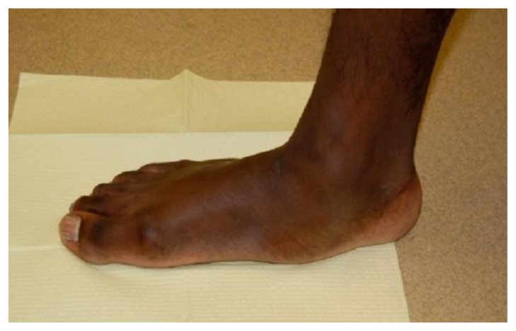
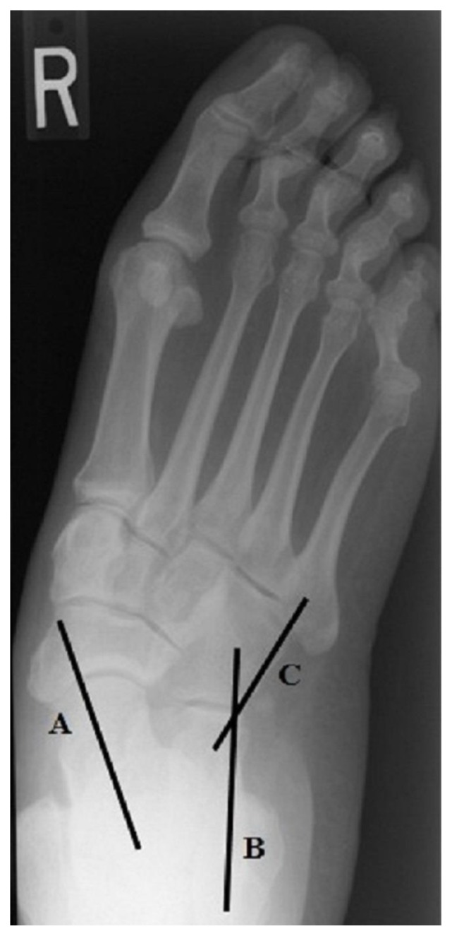
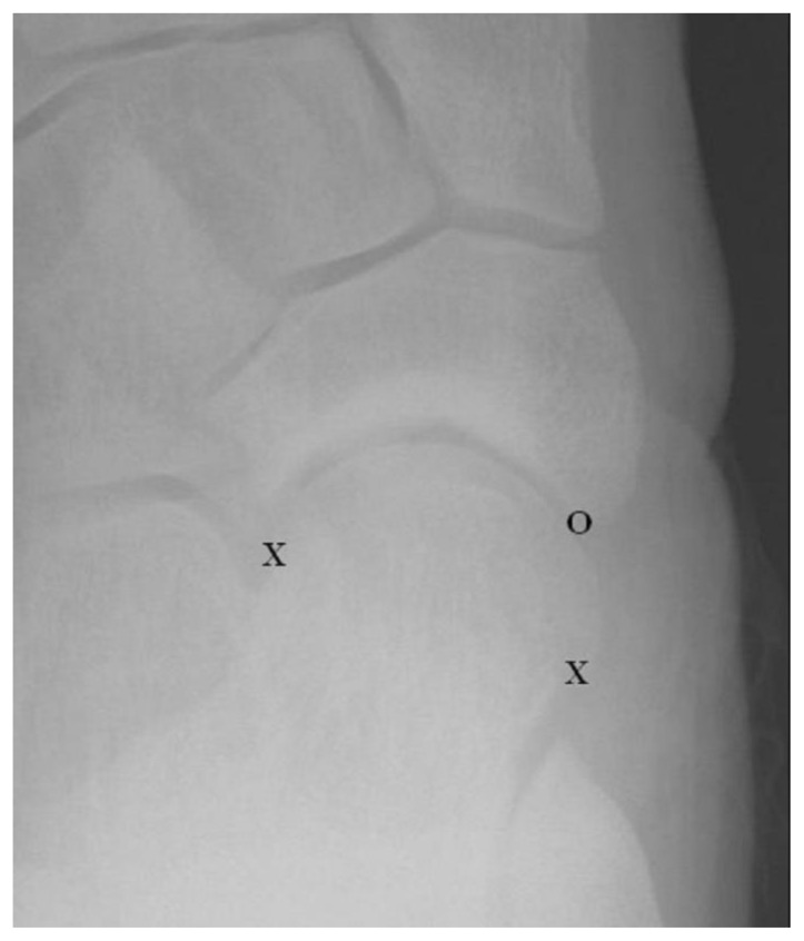
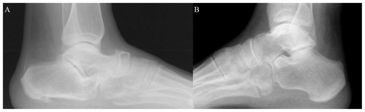
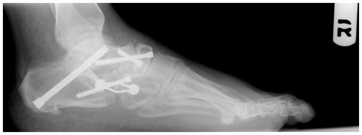
This pictorial review focuses on basic procedures performed within the field of podiatric surgery, specifically for elective reconstruction of the midfoot and rearfoot with focus on the flatfoot deformity. Our goal is to demonstrate objective radiographic parameters that surgeons utilize to initially define the deformity, lead to procedure selection, and judge post-operative outcomes. We hope that radiologists will employ this information to improve their assessment of post-operative radiographs following reconstructive foot surgeries. First, relevant radiographic measurements are defined and their role in procedure selection explained. Second, the specific surgical procedures of the Evans calcaneal osteotomy, medial calcaneal slide osteotomy, Cotton osteotomy, subtalar joint arthroeresis, and arthrodeses of the rearfoot are described. Finally, specific plain film radiographic findings that judge post-operative outcomes for each procedure are detailed.

Keywords: Calcaneal Osteotomy; Coalition; Cotton osteotomy; Flatfoot; Podiatric surgery.


Figures

Figure 1
Figure 1
Clinical picture of a 46 y/o male with a symptomatic right flatfoot deformity. Although the deformity is most easily defined as a “collapse” of the medial column longitudinal arch as pictured, it is actually a complex deformity involving bone, joint and soft tissue abnormality in the transverse, sagittal and frontal planes. Picture courtesy of Dr. Jason Piraino, DPM.
Figure 2
Figure 2
Weight bearing AP view of a 46 y/o male with a symptomatic flatfoot deformity demonstrating a transverse plane component to the deformity. The talocalcaneal angle is defined as the resultant angulation between the longitudinal axis of the talar head and neck (Line A) and a tangent drawn to the lateral side of the calcaneus (Line B), with a normal angle ranging between 25–40 degrees. The calcaneocuboid angle (CCA) is defined as the resultant angular relationship between a tangent drawn along the lateral side of the cuboid (Line C) and a tangent drawn along the lateral side of the calcaneus (Line B), with a normal angle between 0–5 degrees. Both angles increase with progressive transverse plane flatfoot deformity.
Figure 3
Figure 3
Cropped AP view of a left foot of a 55 y/o female highlighting the talar-navicular joint. Another measure of transverse plane alignment is the talar head coverage which estimates what percentage of the relativley convex talar head is “covered” by the relativley concave proximal navicular. Normally approximately 75–100% of the talar head articular surface would be expected to be covered by the articular surface of the proximal navicular, with less coverage noted with progressive transverse plane deformity. The image above designates the most medial and lateral aspects of the cartilage on the talar head with “X”s and the most medial aspect of the navicular indicated with an “O”. Only approximately 60% of the talar head articular cartilage is “covered” by the articular cartilage of the proximal navicular in this image.
Figure 4
Figure 4
Weight bearing lateral view of a 46 y/o male with a symptomatic flatfoot deformity indicating sagittal plane component of the deformity. The talar declination angle is the resultant angulation between the supporting surface (Line A) and the longitudinal axis of the talar head and neck (Line B). The calcaneal inclination is the resultant angulation between the supporting surface (Line A) and a line tangential to the plantar aspect of the calcaneus (Line C). The talo-first metatarsal angle is defined by the angular relationship between the longitudinal bisection of the first metatarsal (Line D) and the longitudinal axis of the talar head and neck (Line B).
Figure 5
Figure 5
These cropped lateral radiographic views highlight differences appreciated in the appearance of the sinus tarsi between flatfeet and cavus feet. The sinus tarsi of the subtalar joint in Figure 5A of a 60 y/o male is not well visualized and may be termed “obliterated” when described in a flatfoot as the lateral talar process fills the space, while the sinus tarsi in Figure 5B of a 32 y/o male is very well visualized and may be termed “bullet hole” when describing a cavus foot.
Figure 6
Figure 6
These cropped lateral radiographic views highlight assessment of the midtarsal or Chopart’s joint. In a rectus, or normally aligned, foot (Figure 6A of a 56 y/o female) one may expect to see a relative “S” shape formed by the outline of the calcaneocuboid and talonavicular joints. This continuous line is also referred to as the Cyma line. In a flatfoot (Figure 6B of a 42 y/o male), this “S” shape is displaced with the talonavicular joint found relatively anterior to the calcaneocuboid joint. This may be described as an “anterior break” in the Cyma line.
Figure 7
Figure 7
This cropped lateral view of a 29 y/o male highlights the medial column of the foot with an associated “break” or “fault” in the navicular-cuneiform joint. One can appreciate a relative depression along the dorsal aspect of the bones (highlighted with the overlying white lines) at the level of the “fault” at the navicular-cuneiform joint.
Figure 8
Figure 8
These long leg calcaneal axial views demonstrate a relatively rectus (Figure 8A of a 32 y/o female) and valgused (Figure 8B of a 46 y/o female) rearfoot. On the left one can appreciate a relative parallel relationship between the long axis of the tibia and the calcaneus compared to the valgused orientation of the calcaneal axis on the right. Further, the tibial axis can be extended to see where the calcaneal tuber strikes the ground relative to this landmark. On the left one can appreciate that the calcaneal tuber strikes the ground in close proximity to the extension of the tibial axis, whereas the calcaneal tuber strikes the ground well lateral to this line on the right.
Figure 9
Figure 9
These cropped lateral and calcaneal axial views of a right foot of a 48 y/o male demonstrate a posterior facet subtalar joint coalition which may result in a rigid flatfoot deformity. On the lateral view (Figure 9A), one can appreciate extra radiodensity extending along the posterior and superior aspects of the subtalar joint (black arrow) which may be referred to as a “halo sign” and suspicious for coalition. This finding is confirmed with the calcaneal axial view (Figure 9B) which demonstrates a lack of joint space at the posterior facet (black arrow).
Figure 10
Figure 10
This lateral radiograph of a 26 y/o male is suspicious for a calcaneal-navicular coalition secondary to an elongated anterior process of the calcaneus (arrow) commonly referred to as an “anteater sign.”
Figure 11
Figure 11
These bone models demonstrate performance of the Evans calcaneal osteotomy. First a sagittal saw or osteotome is used to cut through the anterior aspect of the calcaneus approximately 1cm proximal to the calcaneal-cuboid articulation from lateral to medial (left). Then a wedge of bone graft approximately 1cm in diameter is inserted into the osteotomy effectively “lengthening” the calcaneus (right).
Figure 12
Figure 12
These cropped AP (Figure 12A) and Lateral views (Figure 12B) of a 46 y/o male demonstrate a patient following an Evans calcaneal osteotomy and Cotton medial cuneiform osteotomy (among other procedures). One can appreciate the location of the osteotomies and the presence of bone graft as highlighted by the arrows in the anterior calcaneus and medial cuneiform. An anchor in the navicular is also visible indicating a posterior tibial tendon advancement or flexor digitorum longus tendon transfer.
Figure 13
Figure 13
The primary surgical intervention for correction of a sagittal plane component to a flatfoot deformity is soft tissue in nature and in the form of either an Achilles tendon lengthening (pictured here) or gastrocnmeius recession. These are completely soft tissue in nature, and therefore, not visible on plain film radiographs. Picture courtesy of Dr. John Steinberg, DPM.
Figure 14
Figure 14
These bone models demonstrate performance of the Cotton medial cuneiform osteotomy. First a sagittal saw or osteotome is used to cut the dorsal aspect of the medial cuneiform from dorsal to plantar in line with the first metatarsal-medial cuneiform articulation (Figure 14A). Then a wedge of bone graft is inserted into the osteotomy effectively plantarflexing the first metatarsal (Figure 14B).
Figure 15
Figure 15
This calcaneal axial view of a 46 y/o male demonstrates a medial calcaneal slide osteotomy used for correction of a frontal plane component to a flatfoot deformity. A through-and-through osteotomy is performed through the calcaneal tuber with medial translation of the posterior fragment. This is nearly always held in place with internal fixation. This osteotomy will literally pull the calcaneal axis out of a valgus orientation and the tuber closer to an extension of the tibial axis extension (see Figure 8).
Figure 16
Figure 16
This lateral view of a 13 y/o male of a post-operative patient demonstrates implantation of a subtalar joint arthroeresis within the sinus tarsi. This is primary used on pediatric patients and provides a “stop” to excession subtalar joint motion in the direction of pronation.
Figure 17
Figure 17
This post-operative lateral radiograph of a 42 y/o male demonstrates the so-called “triple arthrodesis,” or fusion of the subtalar, calcaneal-cuboid, and talar-navicular joints of the rearfoot. These joints can be fused individually for correction of deformity, or as a unit as demonstrated here. The joints are surgically prepared in such a way as to correct for triplanar angular deformity, and help in place with multiple internal fixation options.

Similar articles

Cited by

References

    1. Mahan KT, Flanigan KP. Pes plano valgus deformity. In: Banks AS, Downey MS, Martin DE, Miller SJ, editors. McGlamry’s Comprehensive Textbook of Foot and Ankle Surgery. Third edition. Philadelphia: Lippincott, Williams and Wilkins; 2001. pp. 799–861. ASIN: B008GE9GEI.
    1. Dimmick S, Chhabra A, Grujic L, Linklater JM. Acquired flat foot deformity: Postoperative imaging. Semin Musculoskelet Radiol. 2012;16:217–232. - PubMed
    1. Sanner WH. Foot segmental relationships and bone morphology. In: Christman RA, editor. Foot and Ankle Radiology. First edition. ISBN-13. St Louis: Churchill Livingstone; 2003. pp. 272–302.pp. 978–0443087820.
    1. Meyr AJ, Wagoner MR. Descriptive quantitative analysis of rearfoot alignment radiographic parameters. J Foot Ankle Surg. 2015;54(5):860–71. - PubMed
    1. Williamson ERC, Chan JY, Burket JC, Deland JT, Ellis SJ. New radiographic parameter assessing hindfoot alignment in stage II adult-acquired flatfoot deformity. Foot Ankle Int. 2014;36(4):417–423. - PubMed