

Case Reports
doi: 10.3941/jrcr.v11i7.3147.
eCollection 2017 Jul.
A Case Report of Mikulicz Syndrome
Affiliations
- PMID: 29299096
- PMCID: PMC5743147
- DOI: 10.3941/jrcr.v11i7.3147
Item in Clipboard
Case Reports
A Case Report of Mikulicz Syndrome
J Radiol Case Rep.
.
Abstract
Mikulicz Syndrome (MS) is a rare chronic condition characterized by the abnormal enlargement of glandular tissue in the head and neck. Patients usually present with enlarged lacrimal and parotid glands. While this can be a benign self-limiting condition, other complex systemic diseases, such as sarcoidosis, may represent other underlying etiologies. We present a case of MS in a patient with a history of Crohn's disease.
Keywords: Immunoglobulin G; Mikulicz’ Disease; Salivary Gland Diseases; Sjogren’s Syndrome; Sjögren-Mikulicz syndrome; Xerostomia.
Figures

A 43-year-old female with Mikulicz Syndrome. Findings: A frontal picture of the patient demonstrates asymmetric facial swelling in the right parotid region (short thick arrow). The left face and parotid region is normal (thin arrow).

A 43-year-old female with Mikulicz Syndrome. Findings: A preoperative photo from the operating room demonstrates the right facial and parotid swelling (thick arrow).
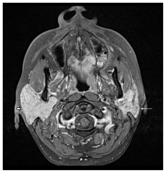
A 43-year-old female with Mikulicz Syndrome. Findings: Axial T1 fat saturated sequence with intravenous contrast demonstrating diffuse enlargement of the right parotid gland (thick short arrow) with homogeneous and normal signal intensity in the right parotid gland without a focal parotid lesion. The left parotid gland is normal in size and signal intensity (thin long arrow). Cranial nerves 5 and 7 were unremarkable (not shown). Technique: 1.5 Tesla magnet, Axial T1 fat saturated sequence with intravenous contrast in venous phase, TR 768ms TE 11ms, gadodiamide 11 mL.

A 43-year-old female with Mikulicz Syndrome. Findings: Axial T2 sequence without contrast demonstrating slightly hypointense T2 signal in the right parotid gland with diffuse enlargement of the right parotid gland (thick short arrow) without a focal parotid lesion. The left parotid gland is normal in size and signal intensity (thin long arrow). Cranial nerves 5 and 7 were unremarkable (not shown). Technique: 1.5 Tesla magnet, Axial T2 sequence without contrast, TR 4340ms TE 113ms.

A 43-year-old female with Mikulicz Syndrome. Findings: Axial STIR sequence without contrast demonstrating diffuse enlargement of the right parotid gland (thick short arrow) with normal and homogeneous signal intensity without a focal parotid lesion. The left parotid gland is normal in size and signal intensity (thin long arrow). Cranial nerves 5 and 7 were unremarkable (not shown). Technique: 1.5 Tesla magnet, Axial STIR sequence without contrast, TR 5930ms TE 87ms TI 140ms.

A 43-year-old female with Mikulicz Syndrome. Findings: Coronal T1 fat saturated sequence with IV contrast demonstrating diffuse enlargement of the right parotid gland (short thick arrow) with homogeneous and normal signal intensity without a focal parotid lesion. The left parotid gland is normal in size and signal intensity (thin long arrow). Cranial nerves 5 and 7 were unremarkable (not shown). Technique: 1.5 Tesla magnet, Coronal T1 fat saturated sequence with IV contrast in venous phase, TR 673ms TE 10ms, gadodiamide 11mL.
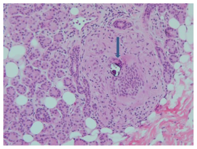
A 43-year-old female with Mikulicz Syndrome. Findings: Hematoxylin and eosin stained pathology section (50× magnification) demonstrates a multinucleated giant cell of the granuloma with a Schaumann body. Schaumann bodies, although not diagnostic, characterize sarcoidosis and are easily seen in the multinucleated giant cells of the granuloma (arrow).
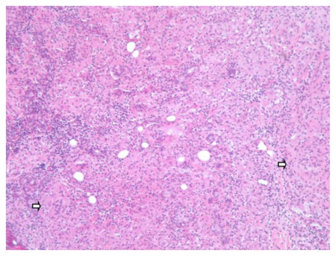
A 43-year-old female with Mikulicz Syndrome. Findings: The hematoxylin and eosin stained pathology section (100× magnification) shows atrophic parotid gland infiltrated sparsely with lymphocytes. Most prominently is the presence of non-necrotizing granulomatosis that are dispersed extensively throughout, predominantly in a micronodular configuration (arrows). Focal confluence of the nodules is evident, which features the sarcoidosis essence of the lesion. Special stains for fungal organisms and mycobacteria were negative (not shown).
References
-
- Mikulicz J. Über eine eigenartige symmetrishe Erkrankung der Thränen-und Mundspeicheldrüsen. In: Mikulicz J, editor. Beiträge zur Chirurgie: Festschrift gewidmet Theodor Billroth. Stuttgart: F.Enke; 1892. pp. 610–630. Available at: http://webapp.uibk.ac.at/alo_cat/card.jsp?id=8542696&pos=54&phys=#.
-
- Schaffer A, Jacobsen A. Mikulicz’s syndrome: A report of ten cases. Am J Dis Child. 1927;34:327–346. DOI: https//doi.org/10.1001/archpedi.1927.04130210002001. - DOI
-
- Napp 0. Ueber die beziehungen der mikuliczschen erkrankung zur tuberculose. Ztschr f Augenh. 1907. pp. 513–517. DOI: https://doi.org/10.1159/000291235. - DOI
-
- Thursfield H. Bilateral salivary swellings (Mikulicz’s disease): A clinical review. QJM. 1914;7:237–249. Available at: http://www.worldcat.org/title/quarterly-journal-of-medicine/oclc/1763235.
Publication types
MeSH terms
LinkOut - more resources
Full Text Sources
Other Literature Sources
Medical