Case Reports
. 2017 Jul 31;11(7):8-13.
doi: 10.3941/jrcr.v11i7.2905. eCollection 2017 Jul.

Paravertebral calcification as a potential indicator for nonaccidental trauma

Affiliations
Case Reports

Paravertebral calcification as a potential indicator for nonaccidental trauma

Katsuaki Kojima et al. J Radiol Case Rep. .

Abstract

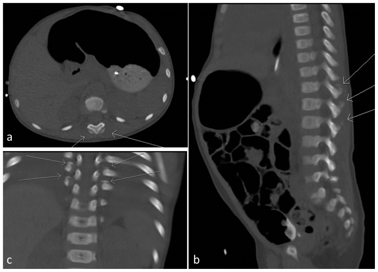
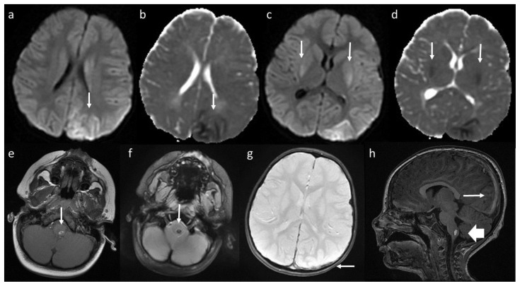
We report a case of nonaccidental trauma (NAT) involving a 23-month-old boy who presented with seizures, acute subarachnoid hemorrhage, and acute subdural hemorrhage. Ophthalmologic examination showed bilateral intraretinal hemorrhages. Further evaluation revealed that he had bilateral thoracolumbar paravertebral calcifications. The Children's Protective Services agency was involved in the case. The child was discharged to an inpatient rehabilitation facility. Vertebral fracture associated with paravertebral calcification has been reported as a sign of NAT. This case was unique because our patient had paravertebral calcifications without vertebral fracture. Paravertebral calcification alone could serve as an indicator of NAT.

Keywords: child abuse; paravertebral calcification; seizure; subarachnoid hemorrhage; subdural hemorrhage.


Figures

Figure 1
Figure 1
23-month-old boy with subdural hemorrhage and subarachnoid hemorrhage. Finding: Computed tomography on day 1 of admission. There is a hyperdensity of the posterior falx (white arrow) as well as sulcal effacement of the left posterior parietal lobe (block arrow). Technique: Axial CT, 150 mA, 100 kV, 3 mm slice thickness, no contrast.
Figure 2
Figure 2
23-month-old boy with paravertebral calcification. Finding: Computed tomography (CT) on day 2 of admission. (a) Soft tissue calcification noted posterior to and to the both sides of the spinous processes of the lower thoracic and upper lumbar spine (arrow). (b) Soft tissue calcification noted posterior to the spinous processes at the lower thoracic and upper lumbar spine including T11 through L1 (arrows). Normal alignment of the vertebral bodies without evidence of acute or healing fractures. (c) Linear soft tissue calcifications adjacent to the rib ends at the costovertebral junctions of the lower thoracic and upper lumbar spine (arrow). Technique: Axial CT (a), 93 mA, 100 kV, 0.5 mm slice thickness, no contrast; sagittal CT (b), 93 mA, 100 kV, 5 mm slice thickness, no contrast; coronal CT (c), 93 mA, 100 kV, 5mm slice thickness, no contrast.
Figure 3
Figure 3
23-month-old boy with left subdural hemorrhage and cerebral infarction in the left cerebral cortex and bilateral posterior basal ganglia. Finding: Brain magnetic resonance image (MRI) on day 3 of admission. (a, b) There is an area of restricted diffusion involving mainly the cortex in the left posterior parietal lobe extending into the left occipital lobe (arrow). (c, d) There is an area of restricted diffusion in the posterior basal ganglia bilaterally (arrow). (e, f) Signal loss on gradient echo images noted within the left posterior medulla (arrow). Given the clinical scenario, this probably reflects deoxyhemoglobin from recent injury. (g) Thin left-sided subdural hematoma overlying the left parietal occipital region (arrow). (i) Subdural hematoma extends medially to the falx and tentorium (arrow). Hyperintensity is noted in left medulla (block arrow). Technique: Brain MRI. 3 Tesla magnet, no contrast used. a. DWI, TR 8000, TE 100.8, no contrast; b. ADC, TR 8000, TE 100.8; c. DWI, TR 8000, TE 100.8; d. ADC, TR 8000, TE 100.8; e. axial T1, TR 616.7, TE 20; f. axial T2 MERGE, TR 600, TE 12.21; g. axial T2 MERGE, TR 600, TE 12.21; h. sagittal T1 BRAVO, TR 11.85, TE 4.8.

References

    1. Roaten JB, Partrick DA, Nydam TL, et al. Nonaccidental trauma is a major cause of morbidity and mortality among patients at a regional level 1 pediatric trauma center. Journal of pediatric surgery. 2006;41:2013–2015. - PubMed
    1. Lindberg DM, Beaty B, Juarez-Colunga E, et al. Pediatrics. 2015;136:831–838. - PubMed
    1. Levin TL, Berdon WE, Cassell I, et al. Pediatr Radiol. 2003;33:305–310. - PubMed
    1. Carrion WV, Dormans JP, Drummond DS, et al. Journal of pediatric orthopedics. 1996;16:210–214. - PubMed
    1. King WK, Kiesel EL, Simon HK. Child abuse fatalities: are we missing opportunities for intervention? Pediatric emergency care. 2006;22:211–214. - PubMed

Publication types

MeSH terms

LinkOut - more resources