. 2017 Nov 22;8(66):110257-110272.
doi: 10.18632/oncotarget.22628. eCollection 2017 Dec 15.

Inhibition of YAP function overcomes BRAF inhibitor resistance in melanoma cancer stem cells

Affiliations

Inhibition of YAP function overcomes BRAF inhibitor resistance in melanoma cancer stem cells

Matthew L Fisher et al. Oncotarget. .

Abstract

Treating BRAF inhibitor-resistant melanoma is an important therapeutic goal. Thus, it is important to identify and target mechanisms of resistance to improve therapy. The YAP1 and TAZ proteins of the Hippo signaling pathway are important drivers of cancer cell survival, and are BRAF inhibitor resistant factors in melanoma. We examine the role of YAP1/TAZ in melanoma cancer stem cells (MCS cells). We demonstrate that YAP1, TAZ and TEAD (TEA domain transcription factor) levels are elevated in BRAF inhibitor resistant MCS cells and enhance cell survival, spheroid formation, matrigel invasion and tumor formation. Moreover, increased YAP1, TAZ and TEAD are associated with sustained ERK1/2 activity that is not suppressed by BRAF inhibitor. Xenograft studies show that treating BRAF inhibitor-resistant tumors with verteporfin, an agent that interferes with YAP1 function, reduces YAP1/TAZ level, restores BRAF inhibitor suppression of ERK1/2 signaling and reduces tumor growth. Verteporfin is highly effective as concentrations of verteporfin that do not impact tumor formation restore BRAF inhibitor suppression of tumor formation, suggesting that co-treatment with agents that inhibit YAP1 and BRAF(V600E) may be a viable therapy for cancer stem cell-derived BRAF inhibitor-resistant melanoma.

Keywords: BRAF inhibitor; YAP and TAZ; cancer stem cell; drug resistance; melanoma.


Conflict of interest statement

CONFLICTS OF INTEREST The authors declare no conflicts of interest.

Figures
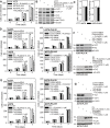
Figure 1
Figure 1. YAP1 and TAZ levels are elevated in A375-PLX-R cells and confer resistance to PLX4032
(A) A375 and A375-PLX-R cells were plated at equal density in growth medium and after overnight attachment were treated from 0 - 3 d with 0 or 1 μM PLX4032. (B) A375 and A375-PLX-R cells, growing in monolayer culture, were treated with 0 or 1 μM PLX4032 for 24 h and extracts were prepared to assay YAP1, TAZ and TEAD level. (C) Cells were grown in monolayer culture and extracts were prepared for qRT-PCR detection of YAP1 and TAZ mRNA. (D, E) A375 and A375-PLX-R cells were double-electroporated with 3 μg of Control-, YAP1- or TAZ-siRNA, or 2 μg of YAP(S127A), TAZ(S89A) or empty vector (EV), and after overnight attachment grown as monolayer cultures in the presence of 0 or 1 μM PLX4032. (F, G) Extracts were prepared from the cells described in panels D/E, at three days of treatment, and assayed by immunoblot to confirm YAP1 and TAZ knockdown and to demonstrate YAP(S127A) and TAZ(S89A) expression. The plotted values are mean ± SEM and asterisks indicate a significant reduction compared to control (n = 3, p < 0.005).
Figure 2
Figure 2. PLX4032 impact on A375 and A375-PLX-R cell spheroid formation and invasion
(A, B) A375 and A375-PLX-R cells were plated in ultra-low attachment plates in spheroid medium, treated with the indicated doses of PLX4032, and spheroid number was monitored. (C) Spheroids were grown for 6 d in the presence of 0 or 1 μM PLX4032 prior to harvest, and lysates were prepared for detection of the indicated epitopes. (D) A375 cells were seeded on chamber slides, treated with 0 or 1 μM PLX4032 for 24 h, then fixed, permeabilized and incubated with primary antibodies specific for the indicated epitope and appropriate secondary antibody (C, control - indicates a staining control where sections were incubated with the secondary antibody alone). (E) A375 and A375-PLX-R cells were seeded atop a matrigel-coated membrane, in growth medium containing 0 or 1 μM PLX4032 for invasion assay. After 20 h, the membrane was removed, rinsed and fixed, and DAPI-stained nuclei were counted on the underside of the membrane. (F, G) A375 and A375-PLX-R cells were double-electroporated with 3 μg of Control-, YAP1- or TAZ-siRNA, or 2 μg of empty (EV), YAP(S127A) or TAZ(S89A) vector and plated atop a matrigel-coated membrane in growth medium containing 0 or 1 μM PLX4032. After 18 h, the membranes were fixed and stained with DAPI to visualize migrated cells. The values are mean ± SEM, n = 3. Asterisks indicate a significant reduction relative to control, p < 0.005.
Figure 3
Figure 3. The role of YAP1, TAZ and TEADs
(A, B) A375 cells were double-electroporated with empty vector (EV), YAP(S127A) or TAZ(S89A) encoding vector, plated and after attachment treated for 24 h with 0 or 1 μM PLX4032. Lysates were then collected for immunoblot. (C) A375 cells were electroporated with each of the indicated constructs and then plated for growth or invasion assays in the presence of 0 or 1 μM PLX4032. For the proliferation study, PLX4032 was added after cell attachment/recovery and cell number was determined at 3 d. For invasion assay, the membranes were fixed, and DAPI stained after 24 h to detect invading cells. The values are mean ± SEM, n = 4. (D, E) A375 or A375-PLX-R cells were double-electroporated with the indicated siRNA and permitted to recover before plating 25,000 cells atop a matrigel-coated membrane for invasion assay in the presence of 0 or 1 μM PLX4032. After 18 h, the membranes were fixed, and DAPI stained to detect invading cells. The values are mean ± SEM, n = 4. Asterisks indicate a significant reduction compared to control, p < 0.005. (F) A375 and A375-PLX-R cells were double-electroporated with pooled siRNA against TEAD1, 2, 3, and 4 (0.75 μg each). At 24 h post-electroporation, the cells were treated with 1 μM PLX4032 for 24 hours and lysates were collected for immunoblot.
Figure 4
Figure 4. Verteporfin suppression of spheroid formation and invasion is associated with reduced YAP1/TAZ level
(A, B) A375 or A375-PLX-R cells (40,000) were seeded in ultra-low attachment plates in spheroid medium, treated with verteporfin, and spheroid number was recorded and images derived at the indicated times. (C) A375 and A375-PLX-R cells were seeded in ultra-low attachment plates in spheroid medium and grown as spheroids for 8 d. Verteporfin treatment was initiated and spheroid number counted after 3 d. (D, E) A375 or A375-PLX-R cells were seeded for invasion assay on a matrigel-coated membrane in growth medium containing 0 - 20 μM verteporfin. After 18 h the membrane was removed and DAPI stained to visualize invading cells on the underside of the membrane. (F) A375 and A375-PLX-R cells were treated for 1 μM verteporfin for 48 h and mRNA extracts were prepared for assay of YAP1 and TAZ mRNA by qRT-PCR. (G, H) A375 and A375-PLX-R cells were plated for spheroid formation followed by addition of 1 μM verteporfin and incubation for 6 d prior to preparation of extracts for immunoblot detection of YAP1, TAZ and pan-TEAD. (I) A375 monolayer cells were pre-treated with 1 μM lactacystin for 1 h followed by addition of 5 μM verteporfin. Lysates were collected after 24 h for immunoblot. The values are mean ± SEM, n = 3 and the asterisks indicate a significant reduction relative to control, p < 0.005.
Figure 5
Figure 5. Verteporfin impact on other PLX4032-resistant melanoma cancer cells
(A) Verteporfin restores sensitivity to PLX4032. A375-PLX-R cells were plated for spheroid formation assay in the presence of the indicated level of PLX4032 and verteporfin which was added the morning after plating. Spheroids were counted at 2, 6 and 10 days. (B) PLX4032-resistant cell lines were harvested, and 40,000 cells were plated in spheroid growth conditions followed by treatment with 0 or 1 μM PLX4032 and counting of spheroid number after 8 d. (C) PLX4032-resistant cell lines were harvested, and 25,000 cells were plated on a matrigel-coated membrane for invasion assay in the presence of 0 or 1 μM PLX4032. After 18 h, the membranes were removed, fixed, and DAPI stained to detect invading cells on the membrane. (D) The indicated melanoma cancer cell lines, maintained in growth medium, were treated for 24 h with 0 or 1 μM PLX4032 and extracts were prepared for immunoblot detection of the indicated proteins. (E) PLX4032-resistant cell lines were harvested, and 40,000 cells were plated in spheroid growth conditions followed by treatment with 0 or 5 μM verteporfin and counting of spheroids after 8 d. (F) PLX4032-resistant cell lines were harvested, and 25,000 cells were plated on a matrigel-coated membrane for invasion assay in the presence of 0 or 5 μM verteporfin. After 18 h, the membranes were removed, fixed, and DAPI stained to detect cells that migrated through the matrigel. (G) SK-MEL-28 cells were double-electroporated with 3 μg of YAP(S127A), TAZ(S89A) or empty (EV) vector and then grown as monolayer cultures in the presence of 0 or 1 μM PLX4032. Cells were counted at the indicated times. (H) PLX4032 resistant A375-PLX-R cells were plated in 35 mm dishes. After 24 h, treatment was initiated with 0 or 1 μM SCH772984 and cells were counted at the indicated times. (I) PLX4032 resistant cells were plated in 35 mm dishes. After 24 h, treatment was initiated with 0 or 1 μM SCH772984 and cells were counted at 3 d. The values are mean ± SEM and asterisks indicate a significant difference compared to control, n = 3, p < 0.005.
Figure 6
Figure 6. Verteporfin impact on tumor formation
(A) Spheroid culture-derived A375 and A375-PLX-R cells were injected at 0.1 million per each front flank and tumor formation was monitored for 4 wks. The asterisk indicates a significant increase in the A375-PLX-R spheroid tumor size compared to the A375 spheroid group, p < 0.005 (B) Spheroid culture-derived A375 and A375-PLX-R cells were injected at 0.1 million cells per each front flank followed by PLX4032 treatment and monitoring of tumor growth for 4 wks. The asterisk indicates a significant reduction in tumor size compared to control group, p < 0.005. (C) Extracts were prepared from A375 and A375-PLX-R tumors (4 wks) from panel B for immunoblot detection of the indicated epitopes. (D, E) A375-PLX-R cells were injected at 0.1 million cells/each front flank and verteporfin treatment was initiated. Extracts were prepared to monitor YAP1 and TAZ levels in control and verteporfin-treated tumors. The asterisk indicates a significant reduction compared to the control group, p < 0.005. (F, G) Spheroid culture-derived A375 and A375-PLX-R cells were grown as tumors for 4 wk with the indicated drug treatment. The tumors were removed and single cell suspensions prepared for matrigel invasion assay. Cells were plated at 25,000 cells per well atop a Matrigel layer in a Millicell chamber in the absence of drug for invasion assay. The labels indicate the drug treatment administered during tumor growth. After 18 h, the membrane was recovered to count migrated cells [38]. The asterisks indicate a significant increase in invasion compared to the A375 spheroid tumor-derived cells, and the double asterisks indicate a reduction in invasion compare to the A375-PLX-R spheroid tumor-derived cells, p < 0.005.
Figure 7
Figure 7. Verteporfin restores PLX4032 suppression of ERK1/2 signaling and tumor formation
(A, B) Spheroid-derived A375-PLX-R cells were injected in each front flank followed by verteporfin and PLX4032 treatment and monitoring of tumor growth for 4 wks. The values are mean ± SEM. The asterisks indicate a significant reduction compared to verteporfin alone, p < 0.001. (C) Tumor extracts were from tumor treated with prepared for detection of signaling proteins. For all figure panels, the asterisks indicate a significant increase compared to control. The values are mean ± SEM, n = 3, p < 0.005.

Comment in

  • Findings of Research Misconduct.
    [No authors listed] [No authors listed] Fed Regist. 2024 Aug 15;89(158):66420-66422. Fed Regist. 2024. PMID: 39161428 Free PMC article. No abstract available.

References

    1. Davies H, Bignell GR, Cox C, Stephens P, Edkins S, Clegg S, Teague J, Woffendin H, Garnett MJ, Bottomley W, Davis N, Dicks E, Ewing R, et al. Mutations of the BRAF gene in human cancer. Nature. 2002;417:949–54. https://doi.org/10.1038/nature00766 - DOI - PubMed
    1. Lito P, Pratilas CA, Joseph EW, Tadi M, Halilovic E, Zubrowski M, Huang A, Wong WL, Callahan MK, Merghoub T, Wolchok JD, de Stanchina E, Chandarlapaty S, et al. Relief of profound feedback inhibition of mitogenic signaling by RAF inhibitors attenuates their activity in BRAFV600E melanomas. Cancer Cell. 2012;22:668–82. https://doi.org/10.1016/j.ccr.2012.10.009 - DOI - PMC - PubMed
    1. Spagnolo F, Ghiorzo P, Queirolo P. Overcoming resistance to BRAF inhibition in BRAF-mutated metastatic melanoma. Oncotarget. 2014;5:10206–21. https://doi.org/10.18632/oncotarget.2602 - DOI - PMC - PubMed
    1. Hauschild A, Grob JJ, Demidov LV, Jouary T, Gutzmer R, Millward M, Rutkowski P, Blank CU, Miller WH, Jr, Kaempgen E, Martín-Algarra S, Karaszewska B, Mauch C, et al. Dabrafenib in BRAF-mutated metastatic melanoma: a multicentre, open-label, phase 3 randomised controlled trial. Lancet. 2012;380:358–65. https://doi.org/10.1016/S0140-6736(12)60868-X - DOI - PubMed
    1. McArthur GA, Chapman PB, Robert C, Larkin J, Haanen JB, Dummer R, Ribas A, Hogg D, Hamid O, Ascierto PA, Garbe C, Testori A, Maio M, et al. Safety and efficacy of vemurafenib in BRAF(V600E) and BRAF(V600K) mutation-positive melanoma (BRIM-3): extended follow-up of a phase 3, randomised, open-label study. Lancet Oncol. 2014;15:323–32. https://doi.org/10.1016/S1470-2045(14)70012-9 - DOI - PMC - PubMed