Review
. 2017 Dec 26;4(4):159-200.
doi: 10.1002/reg2.92. eCollection 2017 Aug.

Mechanisms of urodele limb regeneration

Affiliations
Review

Mechanisms of urodele limb regeneration

David L Stocum. Regeneration (Oxf). .

Abstract

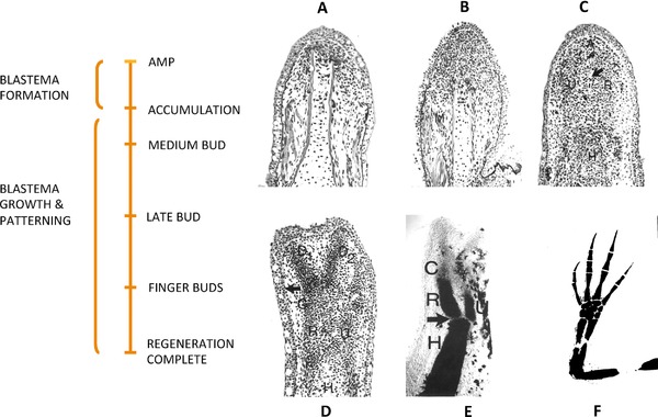
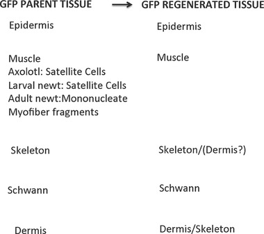
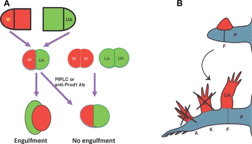
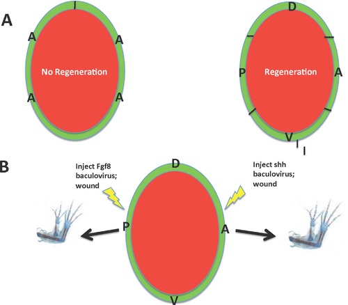
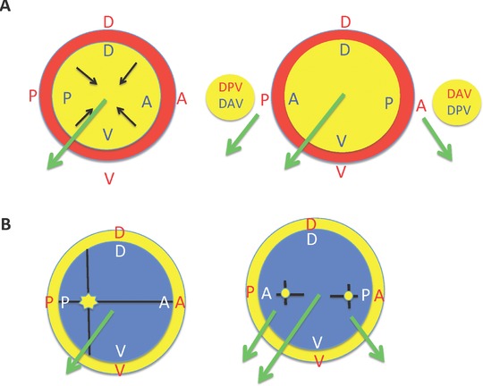
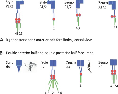
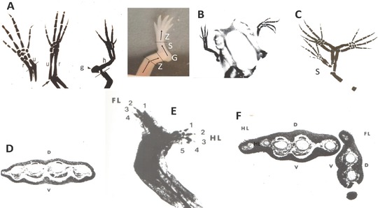
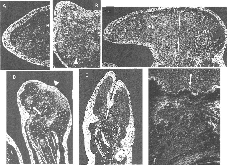
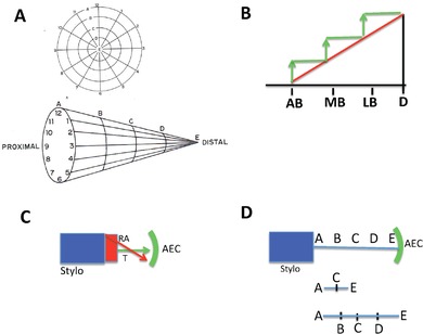
This review explores the historical and current state of our knowledge about urodele limb regeneration. Topics discussed are (1) blastema formation by the proteolytic histolysis of limb tissues to release resident stem cells and mononucleate cells that undergo dedifferentiation, cell cycle entry and accumulation under the apical epidermal cap. (2) The origin, phenotypic memory, and positional memory of blastema cells. (3) The role played by macrophages in the early events of regeneration. (4) The role of neural and AEC factors and interaction between blastema cells in mitosis and distalization. (5) Models of pattern formation based on the results of axial reversal experiments, experiments on the regeneration of half and double half limbs, and experiments using retinoic acid to alter positional identity of blastema cells. (6) Possible mechanisms of distalization during normal and intercalary regeneration. (7) Is pattern formation is a self-organizing property of the blastema or dictated by chemical signals from adjacent tissues? (8) What is the future for regenerating a human limb?

Keywords: limb; mechanisms; regeneration; review; urodele.


Figures

Figure 1
Figure 1
Phases, stages, and longitudinal sections of forelimb regeneration in a urodele larva (Ambystoma maculatum) after amputation through the mid‐stylopodium of the forelimb. Longitudinal sections at various stages of regeneration: (A) accumulation blastema, or early bud; (B) medium bud; (C) late bud, with arrow pointing to a blood vessel, and prominent AER; (D) notch, indicating anlagen of anterior two digits (D1, D2), distal humerus, and radius (R) and ulna (U). The arrow indicates the re‐forming basement membrane. (E) Two‐fingerbud whole mount stained with methylene blue. H, humerus; R, radius; U, ulna; C, carpal region. The arrow points to the elbow joint. (F) Methylene blue stained whole mount of fully regenerated limb. After Stocum (2012, Chapter 8)
Figure 2
Figure 2
Blastema cells have a memory of cellular origin. Results of experiments tracing tissues grafted from transgenic GFP axolotls to white axolotls, based on data from Kragl et al. (2009), Sandoval‐Guzman et al. (2014) and Tanaka et al. (2016). Blastema cells give rise to the same tissue of origin in the regenerate, with the exception of dermal cells, which can also transdifferentiate to skeletal cells. Muscle in larval axolotls and newts is regenerated by satellite cells, whereas regenerated muscle in adult newts is derived primarily from mononucleate cells produced by fragmentation of cut myofibers
Figure 3
Figure 3
Positional memory is encoded in the cell surface. (A) Blastemas derived from the same level (W/W, UA/UA) fuse in a straight line when juxtaposed in culture, but when derived from different levels (W/UA), the more proximal blastema engulfs the distal one. Engulfment is prevented by treating the culture with PIPLC or an antibody to Prod1. (B) Medium bud blastemas derived from the wrist, elbow, and mid upper arm (red) grafted to the dorsal surface of the blastema/stump junction of a hindlimb regenerating from the mid‐femur sort to their corresponding levels of ankle (A), knee (K), and mid‐femur (F) of the regenerating hindlimb. Retinoic acid, which proximalizes the positional identity of blastema cells, abolishes the distal sorting of the wrist and elbow blastemas, so that they behave like upper arm blastema. P, posterior; K, knee; A, ankle; UA, upper arm
Figure 4
Figure 4
(A) Experiment showing that a longitudinal strip of unirradiated skin from one quadrant (here anterior) rotated 90o and grafted as a cuff around the circumference of the amputated internal tissues of an irradiated limb (left) fails to regenerate, but if smaller unirradiated longitudinal strips from each quadrant of the limb (A, anterior; P, posterior; D, dorsal; V, ventral) are rotated and grafted (right), the limb regenerates. (B) Experiment based on Lheureux's model (1977) showing that a baculovirus construct containing the fgf8 gene can substitute for anterior skin and a baculovirus construct containing the shh gene can substitute for posterior skin in evoking supernumerary limb formation at posterior and anterior wound sites on the stylopodium, respectively, to which a nerve has been deviated. S, supernumerary limb
Figure 5
Figure 5
Supernumerary formation. (A) Polar coordinate model. Red, stump; yellow, blastema. Left, normal regeneration. Circumferential fibroblasts interact centripetally (black arrows) to initiate regenerative outgrowth (green arrow). Right, reversal of the blastema AP axis allows interactions between anterior and posterior halves of stump and graft tissues (DPV/DAV, DAV/DPV) to regenerate two supernumerary limbs with stump handedness (shorter green arrows); the graft develops (longer green arrow) with the handedness of origin. (B) Boundary model. Left, normal regeneration. Yellow circle, stump; blue circle, blastema. Interaction between cells at a posteriorly located intersection between AP and DV boundaries triggers the production of a morphogen (star) that initiates regeneration. Right, Reversal of the blastema AP axis creates supernumerary loci of morphogen production (yellow circles) on the anterior and posterior sides of the limb. The primary limb (longer green arrow) has graft handedness and the two supernumerary limbs (shorter green arrows) have stump handedness
Figure 6
Figure 6
Regeneration of anterior and posterior half and double half stylopodia and zeugopodia of axolotl forelimbs. Stylopodium and zeugopodium, blue; carpals, red; digits, green. (A) Half limbs. Posterior and anterior stylopodial halves exhibit non‐equivalent regeneration, with posterior halves regenerating much more than anterior halves, whereas regeneration of posterior and anterior half zeugopodia is more equivalent. (B) Double half limbs. A similar non‐equivalence is exhibited by double anterior stylopodia and zeugopodia
Figure 7
Figure 7
RA‐treated amputated normal limbs, anterior half, dorsal half, double anterior half and double dorsal half limbs with retinoic acid. (A) Normal limb. Left to right, increasing dose of RA after amputation through the distal radius/ulna. r = radius, u = ulna, h = humerus; g = girdle. line = level of amputation. Color photo shows RA‐induced serial PD duplication of hind limb segments after amputation through the distal zeugopodium (Z). G, S, Z indicate the duplicated girdle, stylopodium, and zeugopodium/autopodium. (B) Regeneration from RA‐treated anterior half zeugopodia grafted to the orbit. AP/DV complete, proximalized limbs were regenerated. (C) Regeneration of mirror‐image limb from RA‐treated double anterior zeugopodia. A proximalized supernumerary limb (S) arose where tissue of the grafted anterior half met posterior tissue. (D) Section through a regenerated RA‐treated dorsal half zeugopodium at the level of the distal metacarpals. RA proximalizes these regenerates and ventralizes positional identity, as shown by the normal pattern of extensor muscles on the dorsal side (d) and flexor muscles on the ventral side (v). (E) RA‐treated double dorsal half zeugopodium. One half was fore limb, the other half was hind limb. A fore limb (FL) was regenerated by the forelimb half and a hind limb (HL) by the hindlimb half. Numbers indicate digits. (F) Cross‐section through another such specimen showing that both the forelimb and hind limb regenerated normal DV muscle patterns. After Thoms and Stocum (1984), Kim and Stocum (1986a), and Ludolph et al. (1990)
Figure 8
Figure 8
Histology of RA‐treated limbs. (A) Normal fore limb blastema, no RA treatment. R = radius; U = ulna. (B) RA‐treated medium bud stage blastema, growing posteriorly. Arrowhead indicates proximal high density region of blastema cells. (C) RA‐treated late bud blastema growing perpendicular to the PD axis of the stump. White line, transition between prospective girdle (G) and the free limb (FL). (D) RA‐treated double anterior zeugopodium showing twin condensations of blastema cells separated by thickened epidermis (arrowhead) Arrow points to remnant of zeugopodial stump cartilages. (E) Proximalized twin blastemas growing from a RA‐treated double anterior limb. Arrow indicates basement membrane reforming beneath the epidermis between the blastemas, but not under the rest of the blastemal epidermis. (F) Non‐regenerating RA‐treated double posterior limb with thick basement membrane (arrow) and connective tissue pad under the epidermis. After Kim and Stocum (1986b)
Figure 9
Figure 9
Models of distalization. (A) Polar coordinate model. Top, planar representation. The concentric circles (radial values) labeled A−E represent the PD positional identities generated by successive reiterations of centripetal migration and interaction. Numbers represent angular values. Bottom, the radial values telescoped out as each of the radial values is realized. (B) “Bootstrap” model. Red line, morphogen levels from proximal to distal. Green line, production of an AEC factor that increases morphogen levels in a proximal to distal direction. AB, accumulation blastema; MB, medium bud; LB, late bud; D, digits. (C) Regeneration of the segment of amputation (distal stylopodium, red), driven by a high level of RA (red arrow) that drops off distally to interact with a mitotic timing mechanism to specify remaining PD positional identities. (D) Intercalary averaging mechanism. Missing positional identities represented as A−E. The first step is intercalation of the intermediate positional identity; successive intercalations complete the PD sequence. (A) After French et al. (1976) and Bryant et al. (1981). (D) After Maden (1977)
Figure 10
Figure 10
(A) Normal wrist blastema homografted from a dark axolotl to the double anterior stylopodium of a white animal. The graft regenerated with three forelimb digits (P1−P3) and evoked three supernumerary digits (S1−S3). A symmetrical double femur and tibia was intercalated from the host. Arrow, knee joint. (B) Normal wrist blastema of a white axolotl autografted to the ipsilateral double posterior stylopodium of the hindlimb. The graft formed four forelimb digits (P1−P4) and evoked five supernumerary digits (S1−S5). Primary and supernumerary sets of basipodial elements were regenerated. T5, tarsal 5. Arrow, symmetrical fibulae intercalated from host stump. (C) Double anterior wrist blastema homografted from a dark axolotl to the double anterior stylopodium of a white axolotl. The graft formed two carpals (arrows) and a single digit. A symmetrical distal femur (F) and tibia (T) were intercalated from the host stump. After Stocum (1980b, 1981)
Figure 11
Figure 11
Autonomous development of the blastema. (A) Medium bud blastemal mesenchyme after 21 days of hanging drop culture. The blastema underwent abortive morphogenesis. Arrows point to dark shadows within the cell mass that reflect the development of a primitive cartilage. (B) Medium bud forelimb stylopodial blastema autografted to dorsal fin. (C) Proximal half of a palette stage fore limb stylopodial blastema autografted to the ankle level of the hind limb. The graft dedifferentiated and developed as a fore limb according to its level of origin. T = host tarsals; H = humerus; RU = radius/ulna; C = carpals; D = four digits. (D) Normal fore limb stylopodial blastema homografted to the same level of a double posterior hind limb stylopodium. Arrow = original graft‐host junction. The primary regenerate is forelimb (digits 1−4 on the left) with forelimb basipodial elements and radius/ulna (u). A supernumerary hindlimb regenerated with tibia and fibula (f) and digits 1−5. (E) Medium bud stylopodial blastema homografted from normal limb of a dartk axolotl to the same stylopodial level of the double posterior hind limb of a white axolotl. Large arrow = graft/host junction. The graft developed as a normal forelimb. The graft‐derived tissues suffered chronic immunorejection with dilation of blood vessels and hemostasis (small arrows) that sharply demarcated graft from host tissues. (F) Double anterior hind limb stylopodial blastema homografted distally to a double anterior hind limb zeugopodium. The blastema developed according to its double anterior stylopodial origin, forming a tapered cone of cartilage (arrow). After Stocum (1968a,b), Stocum and Melton (1977), Stocum (1980a), and Stocum (1981)

References

    1. Adams, D. S. , Masi, A. , & Levin, M. (2007). H+ pump‐dependent changes in membrane voltage are an early mechanism necessary and sufficient to induce Xenopus tail regeneration. Development, 134, 1323–1335. - PubMed
    1. Albert, P. , & Boilly, B. (1988). Effect of transferrin on amphibian limb regeneration: a blastema cell culture study. Roux's Archives for Developmental Biology, 197, 193–196. - PubMed
    1. Albert, P. , Boilly, B. , Courty, J. , & Barritault, D. (1987). Stimulation in cell culture of mesenchymal cells of newt limb blastemas by EDGFI or II (basic or acidic FGF). Cell Differentiation, 21, 63–68. - PubMed
    1. Allenby, G. , Bicquel, M. T. , Saunders, M. , Kazmer, S. , Speck, J. , Rosenberger, M. et al. (1993). Retinoic acid receptors and retinoid X receptors: interactions with endogenous retinoic acids. Proceedings of the National Academy of Sciences USA, 90, 30–34. - PMC - PubMed
    1. Anastasia, L. , Sampaolesi, M. , Papini, N. , Oleari, D. , Lamorte, G. , Tringali, C. , … Venerando, B . (2006). Reversine‐treated fibroblasts acquire myogenic competence in vitro and in regenerating skeletal muscle. Cell Death and Differentiation, 13, 2042–2051. - PubMed