. 2017 Dec 6;4(4):227-235.
doi: 10.1002/reg2.93. eCollection 2017 Aug.

Limb regeneration in a direct-developing terrestrial salamander, Bolitoglossa ramosi (Caudata: Plethodontidae): Limb regeneration in plethodontid salamanders

Affiliations

Limb regeneration in a direct-developing terrestrial salamander, Bolitoglossa ramosi (Caudata: Plethodontidae): Limb regeneration in plethodontid salamanders

Claudia Marcela Arenas Gómez et al. Regeneration (Oxf). .

Abstract

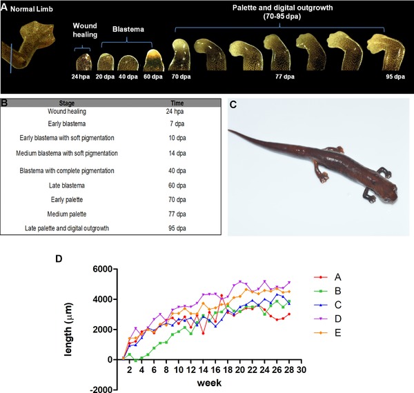
Appendage regeneration is one of the most compelling phenomena in regenerative biology and is extensively studied in axolotls and newts. However, the regenerative capacity in other families of salamanders remains poorly described. Here we characterize the limb regeneration process in Bolitoglossa ramosi, a direct-developing terrestrial salamander of the plethodontid family. We (1) describe the major morphological features at different stages of limb regeneration, (2) show that appendage regeneration in a terrestrial salamander varies from other amphibians and (3) show that limb regeneration in this species is considerably slower than in axolotls and newts (95 days post-amputation for complete regeneration) despite having a significantly smaller genome size than axolotls or newts.

Keywords: axolotl; genome size; newt; plethodontid; urodele.


Figures

Figure 1
Figure 1
Stages of limb regeneration in Bolitoglossa ramosi. (A) The normal limb of B. ramosi is webbed and consists of four digits with an average length of 7.6 ± 1.5 mm (blue line indicates mid‐humerus level plane of amputation). Regenerative stages are indicated: wound healing (24 hpa), blastema (20 dpa), and pigmented blastema (40 dpa), where high pigmentation was noticed, and late blastema (60 dpa). The stages of palette and digital outgrowth occur between 70 and 95 dpa. (B) Nine macroscopic stages were clearly observed, showing that digit extension started after 95 dpa. (C) B. ramosi after 28 weeks post‐amputation (limb with a length of 5.63 ± 1.22 mm). (D) Limb growth rate per day (e.g., [growth in week 2 − growth in week 1]/7 days) in B. ramosi during the 28‐week period, showing the rate for five animals (A, B, C, D, E). hpa, hours post‐amputation; dpa, days post‐amputation
Figure 2
Figure 2
Histological analysis of the adult forelimb in Bolitoglossa ramosi. (A) Staining with Masson's trichrome identifies muscles, connective tissues, and condensation of the cartilage. Additionally, the skin of the distal part of the limb is glandular with high vascularization (V). (B)−(C) Subepithelial glands are abundant surrounding the skin, in particular acinar (yellow arrows) and alveolar glands (green arrows) both with mixed secretions (serous and mucous). (D) The skin at the distal part of the normal limb is highly vascularized (orange arrows) and glandular (yellow arrows). V, vascular tissue; C, cartilage; M, muscle
Figure 3
Figure 3
Histological analysis during limb regeneration in Bolitoglossa ramosi. Hematoxylin and eosin staining at low magnification (A, B, C, D) and high magnification (40×) (A′, B′, C′, D′). (A), (A′) Blastema 3 dpa. A thin wound epidermis covers the regenerate, and inflammatory cells are present (black arrows). (B), (B’) Blastema 20 dpa. The dermal−epidermal junction shows the presence of melanophores and eumelanin. The striated muscle appears fragmented within the regenerate (black arrows in B). Additionally, the differentiating dermis contains immature collagen fibers, and a basement membrane is present (orange arrows in B′). (C), (C′) Blastema 40 dpa. High pigmentation is visible within the blastema as well as the differentiating epithelium. (D), (D’) Early palette 70 dpa. The epidermis is well differentiated, and mesenchymal cells show condensation (orange arrows in D) and differentiation to form the digit primordium. dpa, days post‐amputation. The squares represent the areas of the figures with high magnification
Figure 4
Figure 4
Pigmented blastema at 40 days post‐amputation in Bolitoglossa ramosi. The regenerative epithelium (RE) is thickened with multiple layers. The differentiating dermis shows an accumulation of abundant mature collagen fibers (C) and the limb blastema contains undifferentiated proliferating cells. The regenerating nerves (N) extend into the limb blastema. N, nerve; C, collagen deposition; RE, regenerative epithelium; dpa, days post‐amputation
Figure 5
Figure 5
Cell proliferation in pigmented blastema 40 days post‐amputation during limb regeneration in Bolitoglossa ramosi. (A) Representative image of a blastema showing BrdU immunostaining as a cell proliferation marker; note the very low level of BrdU incorporation in the regenerate. (B) An example of a BrdU‐positive cell (yellow arrows) from another area of a limb regenerate. (C) An example of a eumelanin producing cell (green arrows), which resembles BrdU‐positive cells due to their high pigmentation

References

    1. Al‐Qattan, M. M. , Abd‐Al Wahed, M. M. , Hawary, K. , Alhumidi, A. A. & Shier, M. K. (2014). Recombinant nAG (a salamander‐derived protein) decreases the formation of hypertrophic scarring in the rabbit ear model. BioMed Research International, 2014, 121098. - PMC - PubMed
    1. AmphibiaWeb (2017). AmphibiaWeb: information on amphibian biology and conservation. Available at https://amphibiaweb.org/ (accessed February 24, 2017).
    1. Arenas, C. M. , Gómez‐Molina, A. , & Delgado, J. P. (2015). Maintaining plethodontid salamanders in the laboratory for regeneration studies. Methods in Molecular Biology (Clifton, N.J.), 1290, 71–78. - PubMed
    1. Campbell, L. (2011). Gene expression profile of the regeneration epithelium during axolotl limb regeneration. Developmental Dynamics, 240(7), 1826–1840. - PMC - PubMed
    1. Chippindale, P. T. , Bonett, R. M. , Baldwin, A. S. & Wiens, J. J. (2004). Phylogenetic evidence for a major reversal of life history evolution in plethodontid salamanders. Evolution, 58(12), 2809–2822. - PubMed

LinkOut - more resources