Review
. 2017 Oct 16;2(6):447-452.
doi: 10.1002/lio2.84. eCollection 2017 Dec.

Specimen mapping in head and neck cancer using fluorescence imaging

Affiliations
Review

Specimen mapping in head and neck cancer using fluorescence imaging

Nutte Teraphongphom et al. Laryngoscope Investig Otolaryngol. .

Abstract

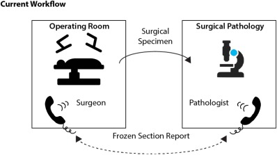
Overview: Although the agreed-upon standard is circumferential pathology analysis of the interface between the resected specimen and the patient, there is currently no consensus on the optimal methodology to achieve this in head and neck cancer specimens. This is most commonly conducted by either sampling the wound bed after resection or obtaining samples from the specimen. Regardless of the technique, only a fraction of the area of interest can be sampled due to the labor-intensive nature of frozen sections.

Objective: This review will cover and define the possible role for optical mapping of the surgical specimen using fluorescence imaging in head and neck cancer.

Level of evidence: NA.

Keywords: Surgery; fluorescence; image‐guidance; oncology; probes.


Figures

Figure 1
Figure 1
(A) Near infrared (NIR) light has the best penetration depth through soft‐tissue. (B) Light when entering a medium can be reflected, scattered and absorbed by molecules within the tissue (chromophores) or excite endogenous or exogenously administered molecules to emit light at a different wavelength.
Figure 2
Figure 2
Cetuximab‐IRDye800 identifies cancer with high specificity. Patient data shown from clinical trial using systemically administered cetuximab‐IRDye800 at 25 mg/m2 3 days prior to surgical excision of head and neck cancer. Patient with oral cavity cancer enrolled for evaluation was noted to have a 5 mm lesion arising within the previous neck dissection scar. (A) Left neck dissection scar, arrow marks 5–6 mm suspicious lesion. (B, C) Intraoperative assessment demonstrated an isolated area of fluorescence within the scar (arrow). (C) The scar underwent wide local excision and repeat LUNA imaging. (D) Ex‐vivo imaging in surgical pathology: specimens were placed into the Pearl Triology system and imaged. (E) Tissue localization of the antibody‐dye bioconjugate in permanent histology sections may assist with disease identification.
Figure 3
Figure 3
Primary tongue specimen from cetuximab‐IRDye800 clinical trial data using microdose. A patient with a lateral tongue squamous cell carcinoma underwent systemic injection of cetuximab‐IRDye800 3 days prior to hemiglossectomy and cervical lymph node dissection. Tongue specimen (A) underwent 4‐mm punch biopsies peripheral (square) and proximal (circle) to the tumor (T). Brightfield imaging (B) and LUNA imaging (C) was performed in the operating room and subsequently imaged in Pathology using the PEARL (D). Histology was used to determine the absence (peripheral biopsy, square) or presence (proximal biopsy, circle) of tumor.
Figure 4
Figure 4
Serial section of tumor and normal muscle by weight from patient treated with anti‐EGFR antibody‐IRDye800. Tumor can be detected as low as 5 mg.
Figure 5
Figure 5
Current workflow. Surgeon sends specimen to pathologist (or leaves the OR to hand carry) with sutures for orientation. A phone conversation required to communicate complex 3D anatomy and areas of suspicion.
Figure 6
Figure 6
Proposed new workflow. Annotation of the suspicious area can be identified and improved overall communication between surgeon and pathologist.

References

    1. McMahon J, O'Brien CJ, Pathak I, et al. Influence of condition of surgical margins on local recurrence and disease‐specific survival in oral and oropharyngeal cancer. Br J Oral Maxillofac Surg 2003;41:224–231. - PubMed
    1. Woolgar JA, Triantafyllou A. Pitfalls and procedures in the histopathological diagnosis of oral and oropharyngeal squamous cell carcinoma and a review of the role of pathology in prognosis. Oral Oncol 2009;45:361–385. - PubMed
    1. Jaafar H. Intra‐operative frozen section consultation: concepts, applications and limitations. Malays J Med Sci MJMS 2006;13:4–12. - PMC - PubMed
    1. Eldeeb H, Macmillan C, Elwell C, Hammod A. The effect of the surgical margins on the outcome of patients with head and neck squamous cell carcinoma: single institution experience. Cancer Biol Med 2012;9:29–33. - PMC - PubMed
    1. Pawlik TM, Scoggins CR, Zorzi D, et al. Effect of surgical margin status on survival and site of recurrence after hepatic resection for colorectal metastases. Ann Surg 2005;241:715–724. - PMC - PubMed