. 2018 Jan 3;3(1):e00545-17.
doi: 10.1128/mSphereDirect.00545-17. eCollection 2018 Jan-Feb.

Microbiota Reconstitution Does Not Cause Bone Loss in Germ-Free Mice

Affiliations

Microbiota Reconstitution Does Not Cause Bone Loss in Germ-Free Mice

Darin Quach et al. mSphere. .

Abstract

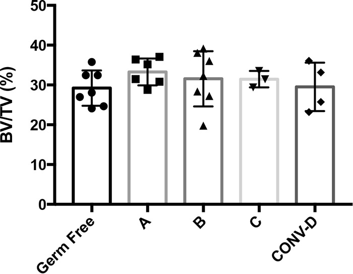
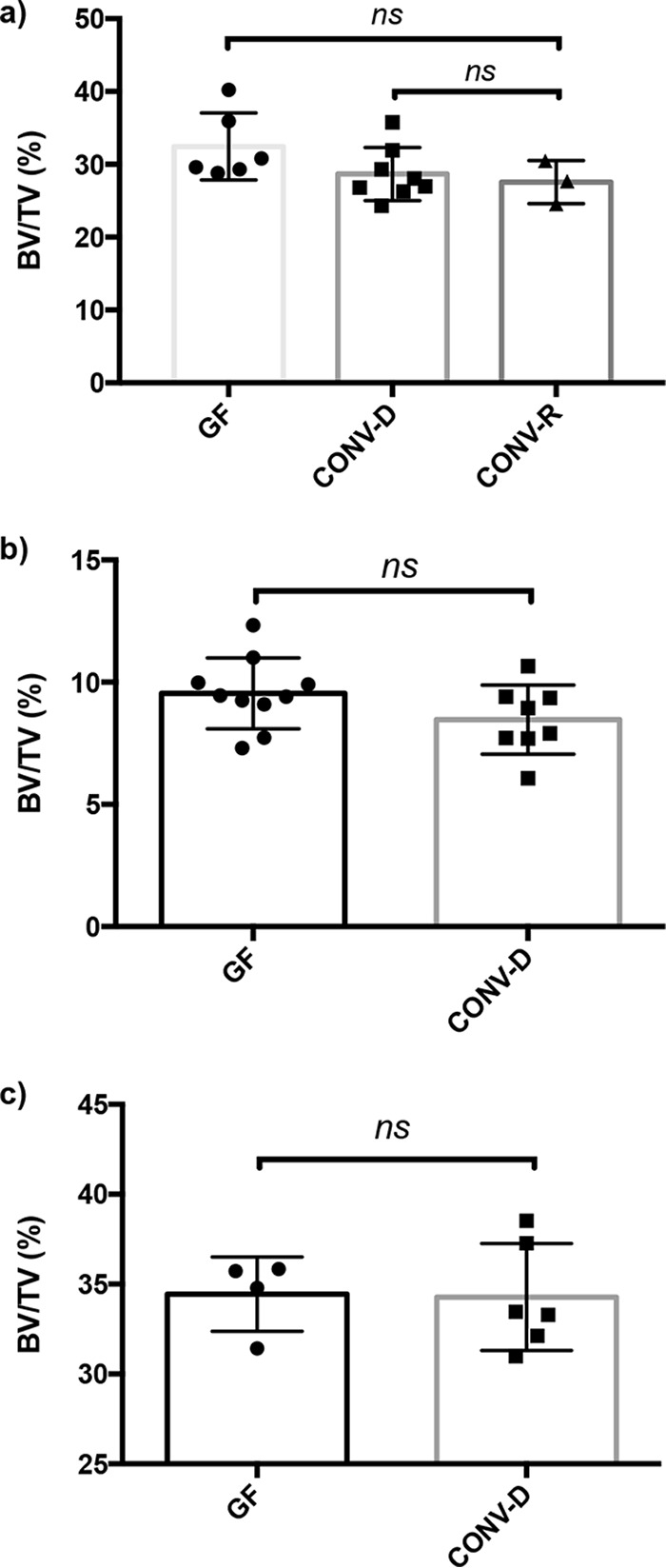
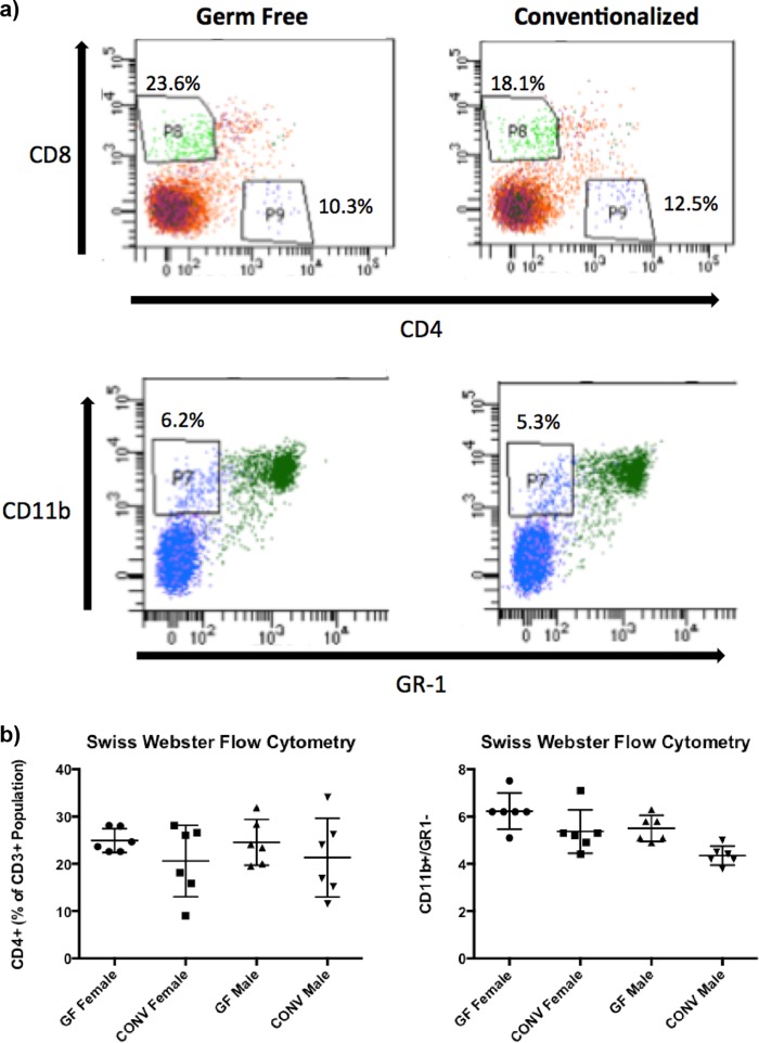
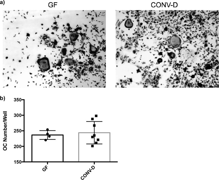
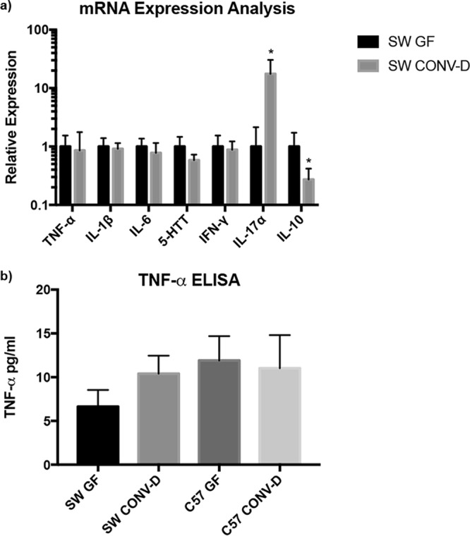
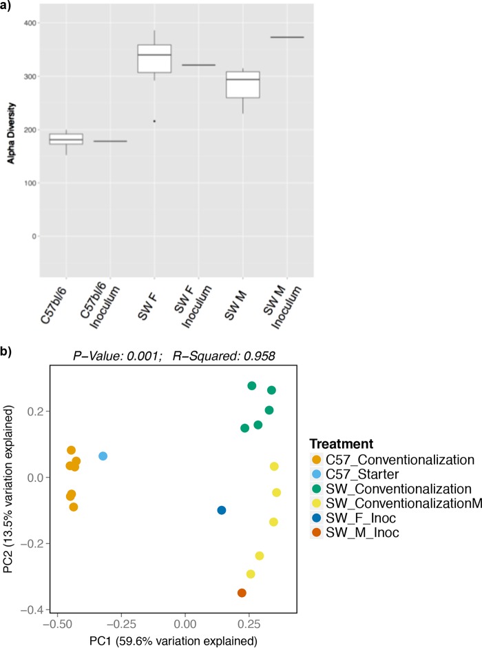
Annually, an estimated 2 million osteoporotic fractures occur in the United States alone. Osteoporosis imparts a great burden on the health care system. The identification of novel regulators of bone health is critical for developing more effective therapeutics. A previous study on the colonization of germ-free (GF) mice with a microbial community has demonstrated that bacterial colonization dramatically increases bone loss. We therefore investigated the impact of multiple microbial communities in different mice to understand how generalizable the impact of bacterial colonization is on bone health. To investigate the impact of different microbial communities on bone health in outbred and inbred mouse strains, gavage was performed on GF Swiss Webster and GF C57BL/6 mice to introduce distinct microbiotas that originated from either humans or mice. GF mice displayed a high degree of colonization, as indicated by more than 90% of the operational taxonomic units present in the starting inoculum being successfully colonized in the mice when they were examined at the end of the experiment. In spite of the successful colonization of GF mice with gut microbiota of either mouse or human origin, bone mass did not change significantly in any of the groups tested. Furthermore, static and dynamic bone parameters and osteoclast precursor and T cell populations, as well as the expression of several inflammatory markers, were mostly unchanged following microbial colonization of GF mice. IMPORTANCE The microbiota has been shown to be an important regulator of health and development. With regard to its effect on bone health, a previous study has suggested that gut microbes negatively impact bone density. However, we show here that this is not generalizable to all microbial communities and mouse strain backgrounds. Our results demonstrate that colonization of mice, both outbred and inbred strains, did not have a major impact on bone health. The identification of microbial communities that do not negatively impact bone health may provide a foundation for future investigations that seek to identify microbes that are either beneficial or detrimental to bone metabolism.

Keywords: bone; microbiome; microbiota; osteoporosis.


Figures

FIG 1
FIG 1
Trabecular bone volume fraction (BVF) values (shown as bone volume per total volume analyzed [BV/TV]) for GF Swiss Webster (SW) mice after colonization with different human microbiotas. Four-week-old female germ-free (GF) Swiss Webster mice were colonized with different human microbiotas (samples A to C) for 8 weeks. The GF and conventionalized (CONV-D) groups served as controls. The results for individual mice and the mean values ± SEM (n = 3 to 7) are shown.
FIG 2
FIG 2
Conventionalization of female GF SW and C57BL/6 mice does not impact bone density. Female GF Swiss Webster (a) and C57BL/6 (b) mice and male GF Swiss Webster (c) mice were colonized with conventional mouse microbiota for 4 weeks (conventionalized [CONV-D]). The GF group served as the control. No differences in BVF were observed when conventionalized mice were compared to their GF counterparts. CONV-R, conventionally reared. The results for individual mice and mean values ± SEM (n = 3 to 10) are shown.
FIG 3
FIG 3
Conventionalization of GF SW mice resulted in no changes in bone marrow cell populations. Bone marrow cells were flushed from 8-week-old mice and stained for antibodies to quantify T cell populations (CD3, CD4, and CD8) and osteoclast precursors (CD11b+/GR1) by flow cytometry. (a) Representative images of flow cytometry plots of cell percentages of total CD3+ population for T cells and total bone marrow population for osteoclast precursors. (b) Quantitation of the different cell populations. The results for individual mice and mean values ± SEM (n = 3 to 10) are shown.
FIG 4
FIG 4
Osteoclast outgrowth from bone marrow cells of GF SW and conventionalized (CONV-D) mice. Bone marrow cells were flushed from the femurs of 8-week-old GF and CONV-D mice and stimulated for differentiation with RANKL and M-CSF. TRAP+ cells were quantitated after 4 days of culture. (a) Representative images of bone marrow cells from mice treated under GF and CONV-D conditions. (b) Total osteoclast (OC) numbers per well were quantitated. The results for individual mice and mean values ± SEM are shown.
FIG 5
FIG 5
Analysis of mRNA expression in the colon and TNF-α levels in serum. (a) The colonic mRNA expression level of IL-17α was upregulated and that of IL-10 was downregulated due to conventionalization. (b) Serum levels of TNF-α were unaffected following conventionalization. ELISA, enzyme-linked immunosorbent assay. *, P < 0.05 with respect to the results for SW GF mice, using Student’s t test.
FIG 6
FIG 6
Alpha and beta diversity measures for conventionalized SW and C57BL/6 mice indicate higher diversity in SW mice and distinct clustering. (a) The comparison of OTUs in starting inocula and conventionalized mice suggests a high level of colonization and efficient microbiota transfer in both SW (male and female) and C57BL/6 mouse genetic background. (b) In a PCoA plot, the results for the conventionalized mice cluster together according to mouse genetic background and starting inocula.

References

    1. Abrams GD, Bauer H, Sprinz H. 1963. Influence of the normal flora on mucosal morphology and cellular renewal in the ileum. A comparison of germ-free and conventional mice. Lab Invest 12:355–364. - PubMed
    1. Bouskra D, Brézillon C, Bérard M, Werts C, Varona R, Boneca IG, Eberl G. 2008. Lymphoid tissue genesis induced by commensals through NOD1 regulates intestinal homeostasis. Nature 456:507–510. doi: 10.1038/nature07450. - DOI - PubMed
    1. Cash HL, Whitham CV, Behrendt CL, Hooper LV. 2006. Symbiotic bacteria direct expression of an intestinal bactericidal lectin. Science 313:1126–1130. doi: 10.1126/science.1127119. - DOI - PMC - PubMed
    1. Maier BR, Hentges DJ. 1972. Experimental Shigella infections in laboratory animals. I. Antagonism by human normal flora components in gnotobiotic mice. Infect Immun 6:168–173. - PMC - PubMed
    1. Fernández-Santoscoy M, Wenzel UA, Yrlid U, Cardell S, Bäckhed F, Wick MJ. 2015. The gut microbiota reduces colonization of the mesenteric lymph nodes and IL-12-independent IFN-γ production during salmonella infection. Front Cell Infect Microbiol 5:93. doi: 10.3389/fcimb.2015.00093. - DOI - PMC - PubMed

LinkOut - more resources