. 2018 Mar 1;159(3):1264-1276.
doi: 10.1210/en.2017-00653.

Sex- and Age-Specific Impact of ERK Loss Within the Pituitary Gonadotrope in Mice

Affiliations

Sex- and Age-Specific Impact of ERK Loss Within the Pituitary Gonadotrope in Mice

Jessica L Brown et al. Endocrinology. .

Erratum in

Abstract

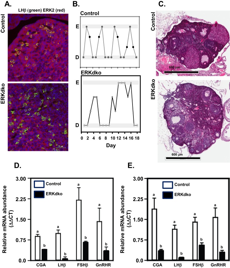
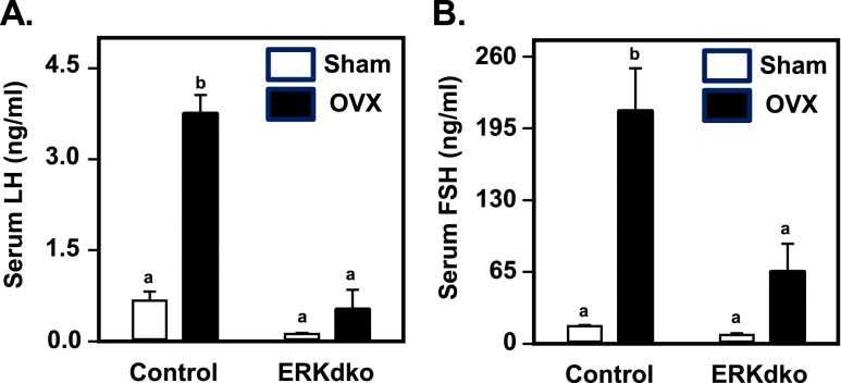
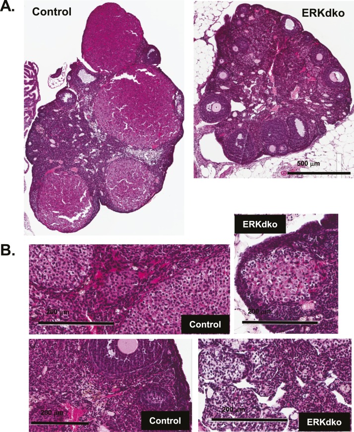
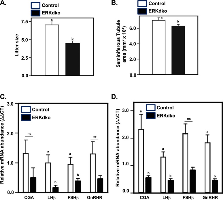
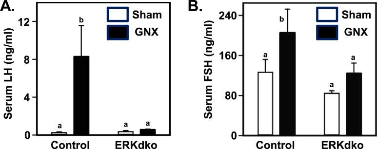
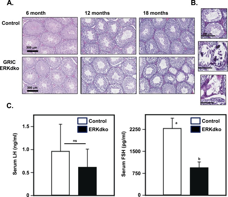
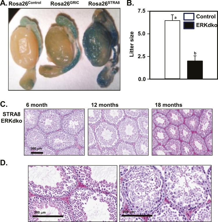
Extracellular signal-regulated kinase (ERK) signaling regulates hormone action in the reproductive axis, but specific mechanisms have yet to be completely elucidated. In the current study, ERK1 null and ERK2 floxed mice were combined with a gonadotropin-releasing hormone receptor (GnRHR)-internal ribosomal entry site-Cre (GRIC) driver. Female ERK double-knockout (ERKdko) animals were hypogonadotropic, resulting in anovulation and complete infertility. Transcript levels of four gonadotrope-specific genes (GnRHR and the three gonadotropin subunits) were reduced in pituitaries at estrus in ERKdko females, and the postcastration response to endogenous GnRH hyperstimulation was blunted. As females aged, they exhibited abnormal ovarian histology, as well as increased body weight. ERKdko males were initially less affected, showing moderate subfertility, up to 6 months of age. Male ERKdko mice also displayed a blunted response to endogenous GnRH following castration. By 12 months of age, ERKdko males had reduced testicular weights and sperm production. By 18 months of age, the ERKdko males displayed reduced testis and seminal vesicle weights, marked seminiferous tubule degeneration, and a 77% reduction in sperm production relative to controls. As the GRIC is also active in the male germ line, we examined the specific role of ERK loss in the testes using the stimulated by retinoic acid 8 (Stra8)-Cre driver. Whereas ERK loss in GRIC and Stra8 males resulted in comparable losses in sperm production, seminiferous tubule histological degeneration was only observed in the GRIC-ERKdko animals. Our data suggest that loss of ERK signaling and hypogonadotropism within the reproductive axis impacts fertility and gonadal aging.


Figures

Figure 1.
Figure 1.
GRIC ERKdko female mice display irregular estrous cycle-like vaginal cytology, hypogonadotropism, and anovulation. (A) Pituitary sections from randomly cycling adult female mice were stained for LHβ (green; to identify gonadotropes) and ERK2 (red). In control animals, overlapping expression of LHβ and ERK2 (yellow) occurred in all cells, whereas in the ERKdko animals, a similar level of overlapping expression was not observed. 4′,6-Diamidino-2-phenylindole nuclear staining is shown in blue. (B) ERKdko animals exhibit irregular periods of estrus and diestrus based on vaginal cytology compared with control animals. (C) Histological sections of ovaries stained with hematoxylin and eosin from control and ERKdko animals. Note the lack of corpora lutea (CL) present in the ovary of ERKdko females. (D) ERKdko animals had substantial reduction in all four gonadotrope-specific gene transcript levels (CGA, LHβ, FSHβ, GnRHR) at 6 months of age compared with control animals (P < 0.05). (E) Gonadotrope-specific transcript levels remained significantly lower in aged ERKdko female mice (12 months) when compared with aged control littermates (P < 0.05). a,bDifferent P < 0.05. D, vaginal cytology at diestrus; E, vaginal cytology at estrus; ΔΔCT, ΔΔ comparative threshold.
Figure 2.
Figure 2.
ERKdko females have a blunted response to GnRH hyperstimulation following ovariectomy. (A) Control animals showed substantial increases in serum LH concentrations following ovariectomy (OVX) compared with sham-operated controls (Sham). In ERKdko animals, basal LH secretion was reduced in sham-operated animals, and response to OVX was blunted. (B) Control animals showed substantial increases in serum FSH concentrations following OVX compared with sham-operated controls (Sham). This effect was blunted in ERKdko female mice. LH and FSH assays were multiplex ELISAs (see Materials and Methods section). a,bDifferent P < 0.05.
Figure 3.
Figure 3.
Aging reveals marked changed in ovarian histology in ERKdko animals. (A) ERKdko animals show abnormal accumulations of extracellular matrix and regions of marked acellularity in histological sections of ovary at 12 months of age. Original magnification, ×500 μm. (B) Higher magnification (×200 μm) of areas of acellularity and extracellular matrix accumulation within the ovaries is depicted.
Figure 4.
Figure 4.
ERKdko males display reduced fertility and are hypogonadotropic. (A) Six-month-old ERKdko males had a significantly reduced litter size compared with control animals. (B) ERKdko males have a significantly reduced seminiferous tubule area compared with control animals at 6 months of age. (C) Six-month-old ERKdko animals have significantly reduced transcript levels of LHβ and FSHβ compared with control animals. Steady-state levels of CGA and GnRHR were numerically smaller but not statistically different (ns) at 6 months of age. (D) Eighteen-month-old ERKdko males had a substantial reduction in CGA, LHβ, and FSHβ transcript levels compared with controls. Steady-state levels of FSHβ mRNA were numerically smaller but not statistically different (ns) at 18 months of age. a,bDifferent P < 0.05.
Figure 5.
Figure 5.
ERKdko males have a blunted response to GnRH hyperstimulation following castration. (A) Control animals showed substantial increases in serum LH concentrations following castration (GNX) compared with sham-operated controls (Sham). In ERKdko animals, the response to GNX was abrogated. (B) Serum FSH response to GNX in control and ERKdko males was numerically higher but not statistically different compared with sham-operated controls (Sham). LH and FSH assays were multiplex ELISAs (see Materials and Methods section). a,bDifferent P < 0.05.
Figure 6.
Figure 6.
Aging reveals marked changed in testicular histology in ERKdko animals. (A) Testes of ERKdko animals showed grossly normal morphology until 18 months of age, where they display signs of testicular degeneration and dysplasia, including regions of marked calcification, aspermatic tubules, and giant spermatid cells. (B) Higher magnification (×200 μm) of regions showing age-related degeneration, calcification, and loss of tubules. (C) LH secretion is numerically lower but not statistically significant (ns) in ERKdko males at 18 months of age. Serum FSH concentrations are reduced (a,bdifferent P < 0.05) in ERKdko males at 18 months of age. LH and FSH assays were in-house assays (see Materials and Methods section).
Figure 7.
Figure 7.
Direct deletion of ERKs in the spermatogonial lineage results in loss of sperm reproduction and fertility but not histological degeneration. (A) The GRIC- and testis-specific Stra8-Cre drivers were compared directly using the Rosa26 β-galactosidase reporter mouse strain. β-Galactosidase staining of whole testes revealed background staining in epididymides in all three genotypes and specific staining within the spermatogonial lineage in the GRIC- and Stra8-Cre strains, albeit in variable staining intensities. (B) Stra8-ERKdko males sire smaller litters compared with control males (a,bdifferent P < 0.05). (C and D) Histological section of testes at 6, 12, and 18 months of age in the Stra8-ERKdko males reveals age-related changes in testicular histopathology, inconsistent with degenerative changes observed in the GRIC ERKdko males at 18 month of age (Fig. 6).

Similar articles

Cited by

References

    1. Bliss SP, Navratil AM, Xie J, Roberson MS. GnRH signaling, the gonadotrope and endocrine control of fertility. Front Neuroendocrinol. 2010;31(3):322–340. - PMC - PubMed
    1. Brown JL, Roberson M. Novel insights into gonadotropin-releasing hormone action in the pituitary gonadotrope. Semin Reprod Med. 2017;35(2):130–138. - PubMed
    1. Clarke IJ, Cummins JT. The temporal relationship between gonadotropin releasing hormone (GnRH) and luteinizing hormone (LH) secretion in ovariectomized ewes. Endocrinology. 1982;111(5):1737–1739. - PubMed
    1. Knobil E. The GnRH pulse generator. Am J Obstet Gynecol. 1990;163(5):1721–1727. - PubMed
    1. Wen S, Schwarz JR, Niculescu D, Dinu C, Bauer CK, Hirdes W, Boehm U. Functional characterization of genetically labeled gonadotropes. Endocrinology. 2008;149(6):2701–2711. - PubMed

Publication types

MeSH terms

Substances