Review
. 2018 Jan 1;19(1):110.
doi: 10.3390/ijms19010110.

The Role of Microglia in Diabetic Retinopathy: Inflammation, Microvasculature Defects and Neurodegeneration

Affiliations
Review

The Role of Microglia in Diabetic Retinopathy: Inflammation, Microvasculature Defects and Neurodegeneration

Christine Altmann et al. Int J Mol Sci. .

Abstract

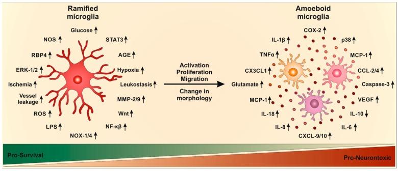
Diabetic retinopathy is a common complication of diabetes mellitus, which appears in one third of all diabetic patients and is a prominent cause of vision loss. First discovered as a microvascular disease, intensive research in the field identified inflammation and neurodegeneration to be part of diabetic retinopathy. Microglia, the resident monocytes of the retina, are activated due to a complex interplay between the different cell types of the retina and diverse pathological pathways. The trigger for developing diabetic retinopathy is diabetes-induced hyperglycemia, accompanied by leukostasis and vascular leakages. Transcriptional changes in activated microglia, mediated via the nuclear factor kappa-light-chain-enhancer of activated B cells (NFκB) and extracellular signal-regulated kinase (ERK) signaling pathways, results in release of various pro-inflammatory mediators, including cytokines, chemokines, caspases and glutamate. Activated microglia additionally increased proliferation and migration. Among other consequences, these changes in microglia severely affected retinal neurons, causing increased apoptosis and subsequent thinning of the nerve fiber layer, resulting in visual loss. New potential therapeutics need to interfere with these diabetic complications even before changes in the retina are diagnosed, to prevent neuronal apoptosis and blindness in patients.

Keywords: angiogenesis; diabetic retinopathy; microglia; neurodegeneration; retina.


Conflict of interest statement

The authors declare no conflict of interest.

Figures

Figure 1
Figure 1
The mouse retina is a model system to analyzing angiogenesis. The mouse retina is a robust tool to analyze in vivo angiogenesis. The avascular retinal vasculature develops gradually as vessels start to grow into the tissue. Starting at postnatal day 0 (P0), blood vessels branch out in a single plane until approximately P8, which allows monitoring of the vessel growth in an intact system including endothelial cells, pericytes, neurons, microglia, astrocytes and Müller cells. Genotype-specific alteration can be analyzed, which may provide insight into molecular and cellular mechanisms of the regulation of angiogenesis. (A) Overview of the retina vasculature stained with the endothelial cell marker platelet endothelial cell adhesion molecule (PECAM; CD31) at P5. The retinal leaves show the branching of the multiple retinal vasculatures. The vessels were stained with (B) PECAM and C ERG to mark endothelial cells and their nuclei, respectively. Scale bar in (A) 500 µm, in (B,C) 250 µm.
Figure 2
Figure 2
Possible mechanisms underlying microglial activation and subsequent inflammatory responses in diabetic retinopathy. Ramified microglia are influenced by hyperglycemia, alterations in ischemia, vessel leakage and upregulation of various mediators. Microglia activation means morphological changes, proliferation and migration. This activation results in an inflammatory response, including downregulation (down arrow) of cytokine IL-10 and growth factors as well as upregulation (up arrow) of various cytokines, chemokines and neurotoxins.

References

    1. Bourne R.R., Stevens G.A., White R.A., Smith J.L., Flaxman S.R., Price H., Jonas J.B., Keeffe J., Leasher J., Naidoo K., et al. Causes of Vision Loss Worldwide, 1990–2010: A Systematic Analysis. Lancet Glob. Health. 2013;1:e339–e349. doi: 10.1016/S2214-109X(13)70113-X. - DOI - PubMed
    1. Fong D.S., Aiello L., Gardner T.W., King G.L., Blankenship G., Cavallerano J.D., Ferris F.L., 3rd, Klein R. Diabetic Retinopathy. Diabetes Care. 2003;26:S99–S102. doi: 10.2337/diacare.26.2007.S99. - DOI - PubMed
    1. Banati R.B., Gehrmann J., Schubert P., Kreutzberg G.W. Cytotoxicity of Microglia. Glia. 1993;7:111–118. doi: 10.1002/glia.440070117. - DOI - PubMed
    1. Fong D.S., Aiello L.P., Ferris F.L., 3rd, Klein R. Diabetic Retinopathy. Diabetes Care. 2004;27:2540–2553. doi: 10.2337/diacare.27.10.2540. - DOI - PubMed
    1. Nentwich M.M., Ulbig M.W. Diabetic Retinopathy—Ocular Complications of Diabetes Mellitus. World J. Diabetes. 2015;6:489–499. doi: 10.4239/wjd.v6.i3.489. - DOI - PMC - PubMed

LinkOut - more resources