

Pancreatic panniculitis: the "bright" side of the moon in solid cancer patients
- PMID: 29301491
- PMCID: PMC5755411
- DOI: 10.1186/s12876-017-0727-1
Pancreatic panniculitis: the "bright" side of the moon in solid cancer patients
Abstract
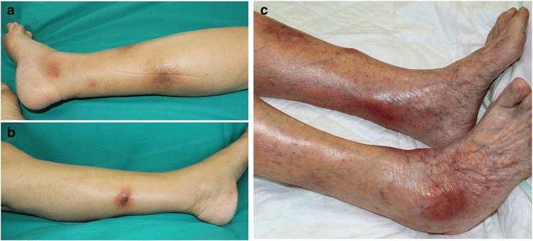
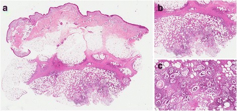
Background: Pancreatic panniculitis is a rare complication of pancreas disorders occurring in 0.3-3% of patients, most often accompanied by the pancreatic acinar carcinoma. It presents multiple, painful, deep, ill-defined, red-brown, migratory nodules and plaques of hard elastic consistency; often ulcerated and typically located on the lower proximal and distal extremities. The pathogenesis is not fully understood, but it is thought to result from lipolysis and fat necrosis with secondary tissue inflammation induced by pancreatic enzymes. Histopathology shows subcutaneous lobular fat necrosis with anuclear adipocytes (called ghost cells) surrounded by a mixed inflammatory infiltrate. Focal calcification may also be seen. The treatment is directed to the underlying disorder, which may result in regression of skin lesions.
Case presentation: We present two cases of pancreatic panniculitis with similar clinical, laboratory, and histopathological features associated with different internal malignancy. The first case, after extensive investigations showed the presence of a pancreatic carcinoma with multiple liver metastases and a poor prognosis. The second one instead is the first case in literature where painful subcutaneous nodules of the legs were the early manifestation of a neuroendocrine carcinoma of the adrenal gland.
Conclusions: Although subcutaneous fat necrosis usually occurs late in the course of a malignancy, recognition of the association with pancreatic panniculitis may prevent a long delay in the diagnosis and management of the occult neoplasm. It should be primarily considered when panniculitis is widespread and persistent, and frequent relapses or tendency to ulcerate of the nodules are regarded as red flags.
Keywords: Pancreatic cancer; Pancreatic panniculitis; Subcutaneous fat necrosis.
Conflict of interest statement
Authors’ information
EG is a resident in Dermatology at the University of Milan, AC1 is a dermatologist, AC2 and RG are dermatologists and pathologists. All of the authors currently work in Italy. AVM is a dermatologist and a former professor at the University of Milan.
Ethics approval and consent to participate
All procedures performed were in accordance with the ethical standards of the institutional and/or national research committee and with the 1964 Helsinki declaration and its later amendments or comparable ethical standards.
Oral informed consent was obtained from the patients for publication of this case report and any accompanying images.
Consent for publication
During lifetime, patients consented orally to the use of their history and all the related images and information for scientific purposes. After the patients’ death the patients’ next of kin gave written consent for the publication of the cases.
Competing interests
The authors declare that they have no competing interests.
Publisher’s Note
Springer Nature remains neutral with regard to jurisdictional claims in published maps and institutional affiliations.
Figures
References
-
- Chiari H. Uber die sogenannte fettnekrose. Prag Med Wochenschr. 1883;8:255–256.
-
- Johnson MA, Kannan DG, Balachandar TG, Jeswanth S, Rajendran S, Surendran R. Acute septal panniculitis. A cutaneous marker of a very early stage of pancreatic panniculitis indicating acute pancreatitis. JOP. 2005;6:334–338. - PubMed
Publication types
MeSH terms
Substances
LinkOut - more resources
Full Text Sources
Other Literature Sources
Medical
