. 2018 Feb;11(2):81-92.
doi: 10.1158/1940-6207.CAPR-17-0267. Epub 2018 Jan 4.

Cyclic-GMP-Elevating Agents Suppress Polyposis in ApcMin mice by Targeting the Preneoplastic Epithelium

Affiliations

Cyclic-GMP-Elevating Agents Suppress Polyposis in ApcMin mice by Targeting the Preneoplastic Epithelium

Sarah K Sharman et al. Cancer Prev Res (Phila). 2018 Feb.

Abstract

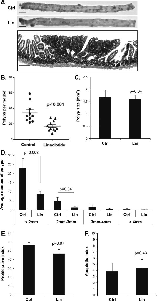
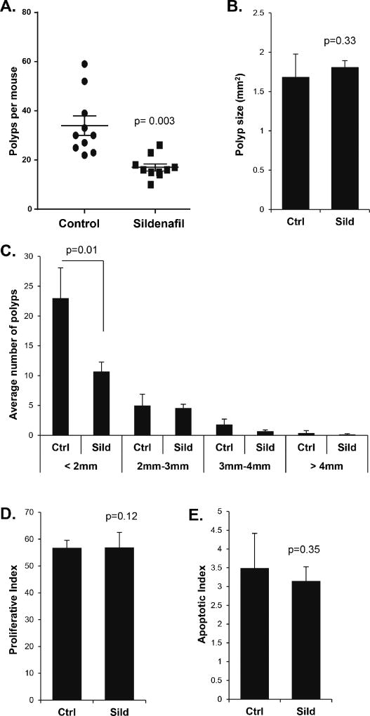
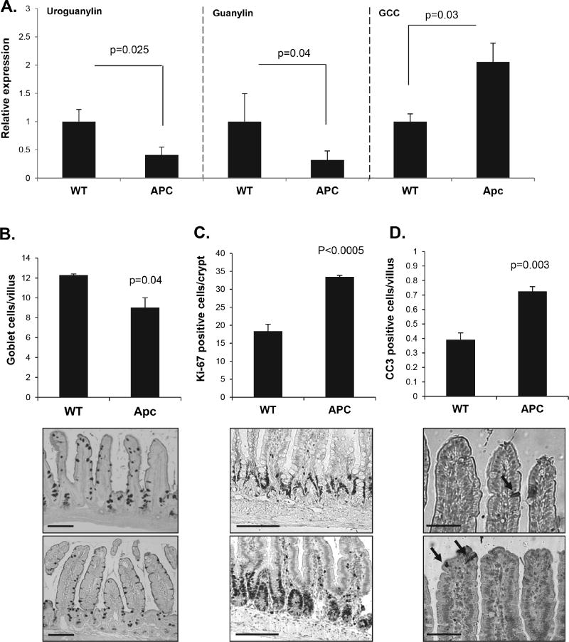
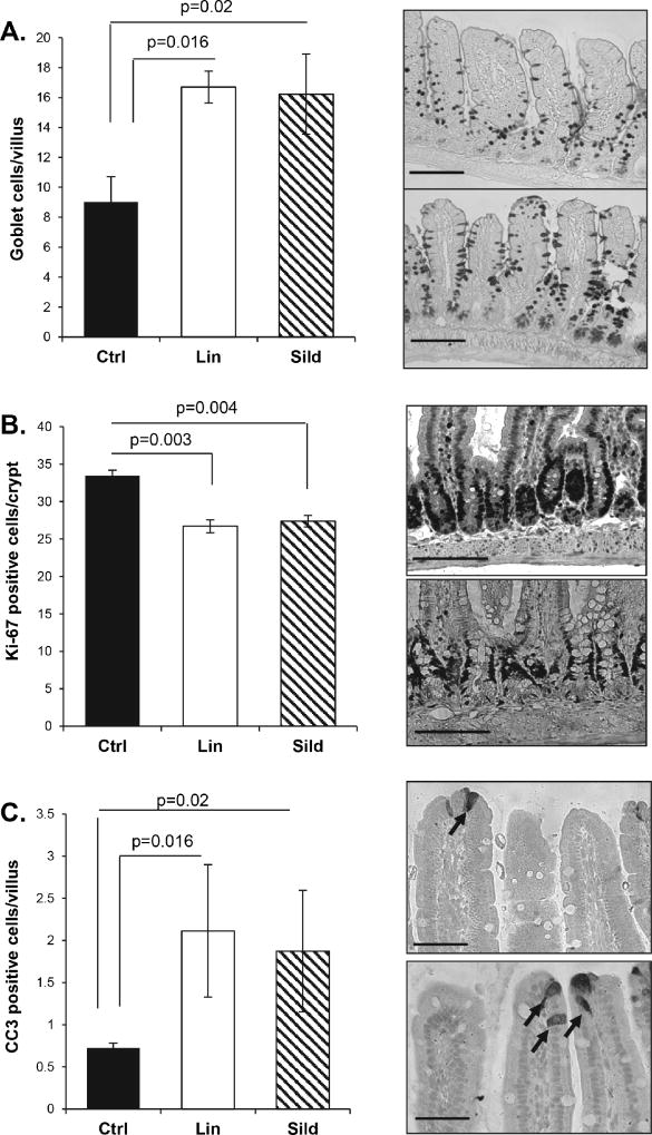
The cGMP signaling axis has been implicated in the suppression of intestinal cancers, but the inhibitory mechanism and the extent to which this pathway can be targeted remains poorly understood. This study has tested the effect of cGMP-elevating agents on tumorigenesis in the ApcMin/+ mouse model of intestinal cancer. Treatment of ApcMin/+ mice with the receptor guanylyl-cyclase C (GCC) agonist linaclotide, or the phosphodiesterase-5 (PDE5) inhibitor sildenafil, significantly reduced the number of polyps per mouse (67% and 50%, respectively). Neither of the drugs affected mean polyp size, or the rates of apoptosis and proliferation. This was possibly due to increased PDE10 expression, as endogenous GCC ligands were not deficient in established polyps. These results indicated that the ability of these drugs to reduce polyp multiplicity was primarily due to an effect on nonneoplastic tissues. In support of this idea, ApcMin/+ mice exhibited reduced levels of endogenous GCC agonists in the nonneoplastic intestinal mucosa compared with wild-type animals, and this was associated with crypt hyperplasia and a loss of goblet cells. Administration of either sildenafil or linaclotide suppressed proliferation, and increased both goblet cell numbers and luminal apoptosis in the intestinal mucosa. Taken together, the results demonstrate that targeting cGMP with either PDE5 inhibitors or GCC agonists alters epithelial homeostasis in a manner that reduces neoplasia, and suggests that this could be a viable chemoprevention strategy for patients at high risk of developing colorectal cancer. Cancer Prev Res; 11(2); 81-92. ©2018 AACR.


Conflict of interest statement

The authors declare no potential conflicts of interest.

Figures

Figure 1
Figure 1. Linaclotide treatment suppresses polyp multiplicity in the ApcMin/+ mouse intestine
A, Upper panels show representative jejunal sections from the intestines of 14-week old ApcMin/+ mice, untreated (Ctrl) or treated for ten-weeks with linaclotide (Lin). Lower panel shows an H&E-stained section containing a representative intestinal polyp from a control animal. B, Plot shows the effect of linaclotide on median polyp number per animal. C, Mean polyp size in ApcMin/+ mice, untreated (Ctrl) or treated for ten-weeks with linaclotide (Lin). D, Average number of polyps in various size classes in ApcMin/+ mice, untreated (Ctrl) or treated for ten-weeks with linaclotide (Lin). E and F, show histological analysis of proliferative index (Ki-67), and apoptotic index (CC3) within polyps from control and linaclotide treated animals. Indices were calculated as staining nuclei (or cells for CC3) as a function of the total number of nuclei stained by hematoxylin. Error bars show SEM, and the p-values are from a two-tailed Student’s t-test. Scale bars represent upper panel 10 mm, lower panel 50 µm.
Figure 2
Figure 2. Sildenafil treatment reduces the number but not the phenotype of intestinal polyps from ApcMin/+ mice
A, The effect of sildenafil on median polyp number per animal. B, Mean polyp size in ApcMin/+ mice, untreated (Ctrl) or treated for ten-weeks with sildenafil (sild). C, Average number of polyps in various size classes in ApcMin/+ mice, untreated (Ctrl) or treated for ten-weeks with sildenafil (Sild). D and E, show histological analysis of proliferative index (Ki-67), and apoptotic index (CC3) within polyps from control and sildenafil-treated animals. Indices were calculated as staining nuclei (or cells for CC3) as a function of the total number of nuclei stained by hematoxylin. Error bars show SEM, and the p-values are from a two-tailed Student’s t-test.
Figure 3
Figure 3. Sildenafil treatment does not reduce inflammation within polyps of ApcMin/+ mice
A, B, C, Real-time qPCR analysis of inflammatory cytokine gene expression in mucosa from C57BL/6 mice and mucosa and polyps from untreated or sildenafil-treated ApcMin/+ mice (n= 6). D, E, Representative images and quantification of Cd11b+ cells (red) in intestines of C57BL/6 mice and untreated or sildenafil-treated ApcMin/+ mice. Tissues were counterstained with DAPI, n= 3 mice, 3 polyps per mouse. Scale bars represent 50 µm.
Figure 4
Figure 4. Expression of cGMP signaling components in intestinal polyps from ApcMin/+ mice
A, The expression of cGMP generators was measured by RT-qPCR in non-neoplastic tissue and untreated and treated polyps from ApcMin/+ mice (Control, Ctrl; Sildenafil, Sild). B, The expression of cGMP effectors were measured by real-time qPCR in non-polyp tissue and polyps from untreated and treated ApcMin/+ mice. C, The expression of phosphodiesterases with activity toward cGMP were measured by RT-qPCR in non-polyp tissue and untreated and treated polyps from ApcMin/+ mice. n= 6 mice; Error bars show SEM, and the p-values are from a One-way ANOVA and Tukey’s post hoc analysis.
Figure 5
Figure 5. Non-polyp epithelium from ApcMin/+ mice appears deficient in cGMP signaling
A, RT-qPCR analysis of uroguanylin, guanylin, and GCC gene expression in C57BL/6 mucosal tissue and non-polyp mucosal tissue from ApcMin/+ mice. B, C, D, Quantification and representative images of goblet cells (AB/PAS), proliferation (Ki-67), and apoptosis (CC3) in stained sections of intestine from C57/BL6 mice and ApcMin/+ mice. A, n=6; B-D, n=3. Error bars show SEM, and p-values are from a two-tailed Student’s t-test. Scale bars represent 50 µm B, C and 25 µm D.
Figure 6
Figure 6. Linaclotide and sildenafil treatment suppress proliferation in the non-polyp epithelium of ApcMin/+ mice
Non-polyp tissue from ApcMin/+ mice treated with sildenafil or linaclotide for ten weeks were collected and prepared for histological analysis. A, B, C, Tissue was stained for goblet cell density (AB/PAS), proliferation (Ki-67), and apoptosis (CC3). Representative pictures of the staining are shown at the right of the graph. Arrows on Panel C indicate CC3 positive cells. Error bars show SEM, and the p-values are from a two-tailed Student’s t-test. Scale bars represent 50 µm A, B and 25 µm C.

References

    1. Siegel RL, Miller KD, Fedewa SA, Ahnen DJ, Meester RGS, Barzi A, et al. Colorectal cancer statistics 2017. CA: A Cancer Journal for Clinicians. 2017;67(3):177–93. doi: 10.3322/caac.21395. - DOI - PubMed
    1. Drew DA, Cao Y, Chan AT. Aspirin and colorectal cancer: the promise of precision chemoprevention. Nat Rev Cancer. 2016;16(3):173–86. doi: 10.1038/nrc.2016.4. - DOI - PMC - PubMed
    1. Gwyn K, Sinicrope FA. Chemoprevention of colorectal cancer. Am J Gastroenterol. 2002;97(1):13–21. - PubMed
    1. Rothwell PM, Fowkes FG, Belch JF, Ogawa H, Warlow CP, Meade TW. Effect of daily aspirin on long-term risk of death due to cancer: analysis of individual patient data from randomised trials. Lancet. 2011;377(9759):31–41. doi: 10.1016/s0140-6736(10)62110-1. - DOI - PubMed
    1. Thompson WJ, Piazza GA, Li H, Liu L, Fetter J, Zhu B, et al. Exisulind induction of apoptosis involves guanosine 3',5'-cyclic monophosphate phosphodiesterase inhibition, protein kinase G activation, and attenuated beta-catenin. Cancer Res. 2000;60(13):3338–42. - PubMed

Publication types

MeSH terms