. 2018 Apr 1;24(7):1654-1666.
doi: 10.1158/1078-0432.CCR-17-2168. Epub 2018 Jan 4.

Establishing a Preclinical Multidisciplinary Board for Brain Tumors

Affiliations

Establishing a Preclinical Multidisciplinary Board for Brain Tumors

Birgit V Nimmervoll et al. Clin Cancer Res. .

Abstract

Purpose: Curing all children with brain tumors will require an understanding of how each subtype responds to conventional treatments and how best to combine existing and novel therapies. It is extremely challenging to acquire this knowledge in the clinic alone, especially among patients with rare tumors. Therefore, we developed a preclinical brain tumor platform to test combinations of conventional and novel therapies in a manner that closely recapitulates clinic trials.Experimental Design: A multidisciplinary team was established to design and conduct neurosurgical, fractionated radiotherapy and chemotherapy studies, alone or in combination, in accurate mouse models of supratentorial ependymoma (SEP) subtypes and choroid plexus carcinoma (CPC). Extensive drug repurposing screens, pharmacokinetic, pharmacodynamic, and efficacy studies were used to triage active compounds for combination preclinical trials with "standard-of-care" surgery and radiotherapy.Results: Mouse models displayed distinct patterns of response to surgery, irradiation, and chemotherapy that varied with tumor subtype. Repurposing screens identified 3-hour infusions of gemcitabine as a relatively nontoxic and efficacious treatment of SEP and CPC. Combination neurosurgery, fractionated irradiation, and gemcitabine proved significantly more effective than surgery and irradiation alone, curing one half of all animals with aggressive forms of SEP.Conclusions: We report a comprehensive preclinical trial platform to assess the therapeutic activity of conventional and novel treatments among rare brain tumor subtypes. It also enables the development of complex, combination treatment regimens that should deliver optimal trial designs for clinical testing. Postirradiation gemcitabine infusion should be tested as new treatments of SEP and CPC. Clin Cancer Res; 24(7); 1654-66. ©2018 AACR.


Conflict of interest statement

Conflicts of interest: none of the authors declare a conflict of interest relating to work reported in this manuscript.

Figures

Figure 1
Figure 1. Composition of the preclinical Multi-Disciplinary Tumor Board and the multistep approach taken to develop new treatment approaches
BBB=blood brain barrier; R=randomization.
Figure 2
Figure 2. Preclinical brain imaging and neurosurgery
A, Concurrent MRI (top) and bioluminescence (bottom) imaging of the same mouse with a SEP-CR(+) tumor. B, Correlation of MRI and bioluminescence imaging of the same cohort of five mice with SEP-CR(+) tumors. C, Preclinical mouse neurosurgical protocol. D–H, (left) bioluminescence measurement of tumor growth. In parenthesis are the days (d) when treated tumor volume was significantly (p<0.05, non-parametric) less than control. (Right) survival curves of mice with indicated animal numbers and tumor types treated with surgery or control. P value=Log Rank relative to control.
Figure 3
Figure 3. Preclinical fractionated surgery and irradiation
A, Preclinical mouse radiation protocol. B–F, (left) bioluminescence measurement of tumor growth. In parenthesis are the days (d) when treated tumor volume was significantly (p<0.05, non-parametric) less than control. (Right) survival curves of mice with indicated tumor types treated with fractionated irradiation or control. G, Preclinical combination mouse surgery and radiotherapy protocol. H–J, Bioluminescence measures of tumor growth (left) and survival curves (right) of mice with the indicated tumor types treated with surgery with or without fractionated irradiation (P values are Log Rank relative to control. Comparisons between treatments are Log Rank not significant (ns), P<0.005(**), P<0.0005(***), P<0.00005(****).
Figure 4
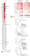
Figure 4. Preclinical repurposing of chemotherapies
A, Repurposing screen of 114 FDA approved and/or clinical trial drugs. Heatmap (left) reports 72 hour IC50 against the indicated cell type; grey bars (middle) indicate FDA approved drugs; graph (right) reports number of completed trials of each drug. Arrows denote drugs selected for further study in (B). B, ‘Washout studies’: heatmaps report IC50 values after timed exposures of the indicated cell types to drug. C–D, (top) bioluminescence measurement of tumor growth. In parenthesis are the days (d) when treated tumor volume was significantly (p<0.05, non-parametric) less than control. (Bottom) survival curves of mice with indicated tumor types treated with the indicated drug monotherapy. Comparisons between treatments are Log Rank not significant (ns) and P<0.0005(***).
Figure 5
Figure 5. Pharmacokinetic, toxicity and efficacy studies of gemcitabine
Plasma and tECF concentration-time plots in the indicated tumor types treated with 60mg/kg bolus (A) or 3 hour infusion (B) gemcitabine. C, Graphs showing induction of tumor cell apoptosis measured by cleaved Caspase 3 immunohistochemistry in mice with the indicated brain tumors treated with 60mg/kg or 3 hour infusion of gemcitabine. D, Toxicity determined by weight loss in mice treated with the indicated doses and regimens of gemcitabine (in C, D: *, p<0.05; **, p<0.005; ***, p<0.0005, Mann-Whitney), E–J, (left) bioluminescence measurement of tumor growth in mice treated with indicated dose and schedule of gemcitabine. In parenthesis are the days (d) when treated tumor volume was significantly (p<0.05, non-parametric) less than control. (Right) survival curves of the same mice shown left. P value=Log Rank relative to control.
Figure 6
Figure 6. Combination surgery, fractionated irradiation, and post-irradiation gemcitabine therapy
Bioluminescence measures of tumor growth (A) and survival curves (B) of mice with mSEP-CR(+) treated with surgery and radiation alone or surgery, radiation and 3 hour infusions of gemcitabine. Bioluminescence measures of tumor growth (C) and survival curves (D) of mice with mCPC treated with surgery and 3 hour infusions of gemcitabine alone or surgery and gemcitabine. Figures in paraenthesis in bioluminescence plots are the days (d) when treated tumor volume was significantly (p<0.05, non-parametric) less than control.

References

    1. Gilbert MR, Sulman EP, Mehta MP. Bevacizumab for newly diagnosed glioblastoma. N Engl J Med. 2014;370:2048–9. - PubMed
    1. Gajjar A, Bowers DC, Karajannis MA, Leary S, Witt H, Gottardo NG. Pediatric Brain Tumors: Innovative Genomic Information Is Transforming the Diagnostic and Clinical Landscape. J Clin Oncol. 2015;33:2986–98. - PMC - PubMed
    1. Brinkman TM, Krasin MJ, Liu W, Armstrong GT, Ojha RP, Sadighi ZS, et al. Long-Term Neurocognitive Functioning and Social Attainment in Adult Survivors of Pediatric CNS Tumors: Results From the St Jude Lifetime Cohort Study. J Clin Oncol. 2016;34:1358–67. - PMC - PubMed
    1. Chemaitilly W, Li Z, Huang S, Ness KK, Clark KL, Green DM, et al. Anterior hypopituitarism in adult survivors of childhood cancers treated with cranial radiotherapy: a report from the St Jude Lifetime Cohort study. J Clin Oncol. 2015;33:492–500. - PMC - PubMed
    1. Krueger DA, Care MM, Holland K, Agricola K, Tudor C, Mangeshkar P, et al. Everolimus for subependymal giant-cell astrocytomas in tuberous sclerosis. N Engl J Med. 2010;363:1801–11. - PubMed

Publication types