. 2018 Feb 16;122(4):568-582.
doi: 10.1161/CIRCRESAHA.117.311240. Epub 2018 Jan 4.

Autophagy Is Required for Sortilin-Mediated Degradation of Apolipoprotein B100

Affiliations

Autophagy Is Required for Sortilin-Mediated Degradation of Apolipoprotein B100

Jaume Amengual et al. Circ Res. .

Abstract

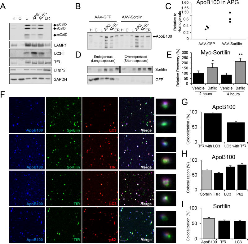
Rationale: Genome-wide association studies identified single-nucleotide polymorphisms near the SORT1 locus strongly associated with decreased plasma LDL-C (low-density lipoprotein cholesterol) levels and protection from atherosclerotic cardiovascular disease and myocardial infarction. The minor allele of the causal SORT1 single-nucleotide polymorphism locus creates a putative C/EBPα (CCAAT/enhancer-binding protein α)-binding site in the SORT1 promoter, thereby increasing in homozygotes sortilin expression by 12-fold in liver, which is rich in this transcription factor. Our previous studies in mice have showed reductions in plasma LDL-C and its principal protein component, apoB (apolipoprotein B) with increased SORT1 expression, and in vitro studies suggested that sortilin promoted the presecretory lysosomal degradation of apoB associated with the LDL precursor, VLDL (very-low-density lipoprotein).

Objective: To determine directly that SORT1 overexpression results in apoB degradation and to identify the mechanisms by which this reduces apoB and VLDL secretion by the liver, thereby contributing to understanding the clinical phenotype of lower LDL-C levels.

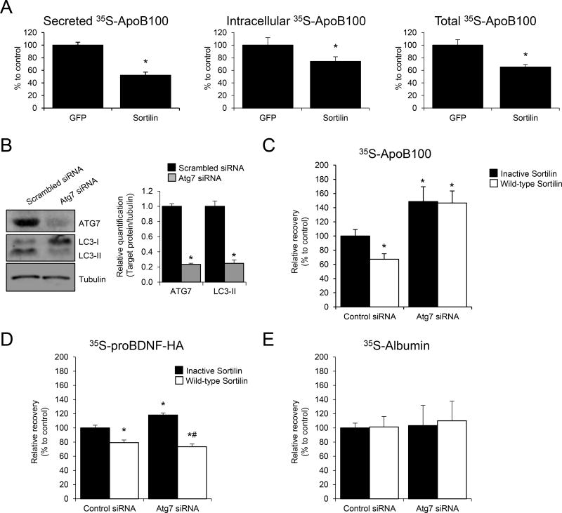
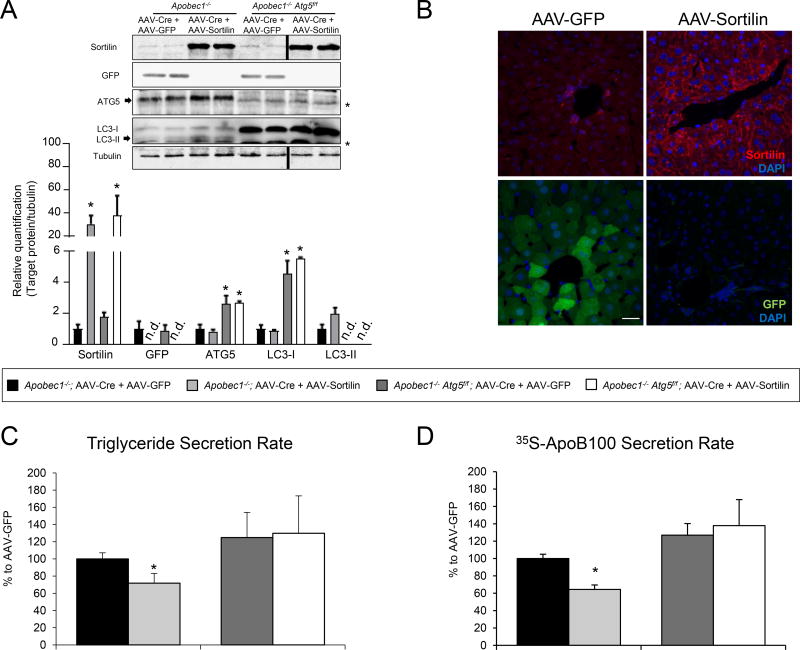
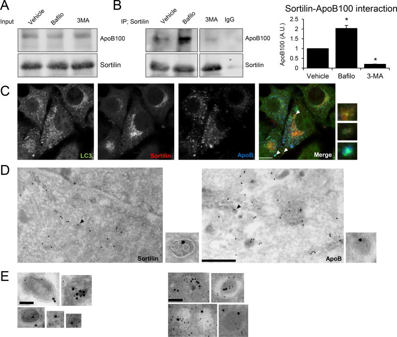
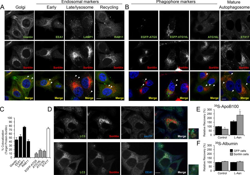
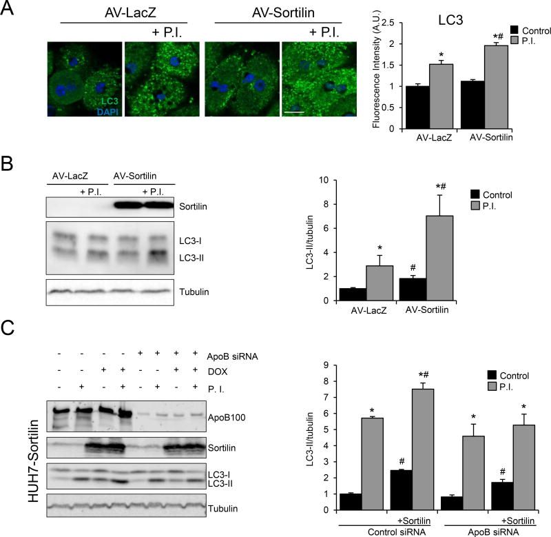
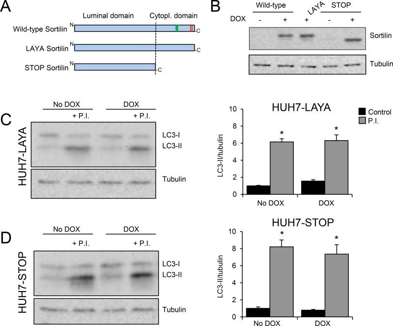
Methods and results: Pulse-chase studies directly established that SORT1 overexpression results in apoB degradation. As noted above, previous work implicated a role for lysosomes in this degradation. Through in vitro and in vivo studies, we now demonstrate that the sortilin-mediated route of apoB to lysosomes is unconventional and intersects with autophagy. Increased expression of sortilin diverts more apoB away from secretion, with both proteins trafficking to the endosomal compartment in vesicles that fuse with autophagosomes to form amphisomes. The amphisomes then merge with lysosomes. Furthermore, we show that sortilin itself is a regulator of autophagy and that its activity is scaled to the level of apoB synthesis.

Conclusions: These results strongly suggest that an unconventional lysosomal targeting process dependent on autophagy degrades apoB that was diverted from the secretory pathway by sortilin and provides a mechanism contributing to the reduced LDL-C found in individuals with SORT1 overexpression.

Keywords: amphisomes; apoB100; autophagy; liver; sortilin.


Figures

Figure 1
Figure 1. Sortilin overexpression increases apoB100 degradation in rat hepatic cells in an autophagy-dependent manner
(A) McA rat hepatic cells stably expressing GFP or sortilin were cultured in normal growth medium. When cells reached sub-confluence (80%–90%), they were washed with PBS, and amino acid-starved for 1 h before being subjected to steady state metabolic labeling using a mixture of [35S]-methionine and cysteine. After 4 h, cells were harvested and apoB100 was immunoprecipitated, separated by SDS-PAGE, and bands quantified using a phosphorimager. The graphical data are displayed as the relative differences in secreted apoB100 (left panel), intracellular apoB100 (middle panel), and total (cell+medium), right panel apoB100 between the cells transfected with GFP (set to 100%) vs. sortilin. (B–E) McA hepatic cells stably expressing human apoB100 were transfected with Atg7 siRNA or with scrambled siRNA for 24 h. Then, cells were transfected with either a plasmid containing the full-length myc-tagged human sortilin (Wild-type Sortilin, pcDNA3.1-hSORT1-myc) or a dominant negative sortilin (“Inactive Sortilin”; pcDNA3.1-hPro-Sortilin) generated by mutation of the furin recognition site, which prevents sortilin binding to apoB100. (B) Western blot for ATG7, LC3-I and LC3-II levels 48 h after siRNA incubation. Tubulin was used as internal control. (C) Pulse-chase results showing the recoveries of apoB100 at the end versus beginning of the chase period, with “Inactive Sortilin, Control siRNA” set to 100%. (D) Relative recovery of proBDNF-HA from McA hepatic cells transfected with Atg7 siRNA or with scrambled siRNA for 24 h. Prior to the pulse-chase study, an expression plasmid for proBDNF-HA was co-transfected with an expression plasmid for either wild-type or inactive sortilin for 24 h. (E) Relative recovery of albumin. Numerical data represent the means ± SEM. *, p<0.05 versus Control siRNA; #, p<0.05 versus Atg7 siRNA using two-tailed Student’s t-test. The experiments were performed in triplicate.
Figure 2
Figure 2. Sortilin-mediated effects on VLDL production are abrogated in liver-specific autophagy deficient mice
Age and sex matched Apobec1−/− and Apobec1−/− Atg5f/f mice received a single intraperitoneal injection containing a combination of two different adeno-associated vectors (AAV) under control of the liver-specific thyroxine-binding globulin promoter. All the mice received Cre recombinase-AAV (AAV-Cre), combined with either GFP or sortilin-containing AAV two weeks before the experiments. (A) Immunoblot analysis of GFP, sortilin, ATG5, LC3 and tubulin in liver homogenates from the indicated mice. * denote non-specific bands. (B) Immunodetection of sortilin (red) and GFP (green) in Apobec1−/− mice injected with AAV-GFP or AAV-Sortilin. DAPI (blue) was used to stain the nuclei. White bar; 25 µm. (C–D) Secretion rates of triglyceride and apoB100 were determined in vivo as described in the Methods in the indicated mice after infection with AAV-GFP (black bars) or AAV-Sortilin (open bars). (C) VLDL-triglyceride secretion rate, and (D) ApoB100 secretion rate. The total amount of plasma proteins radiolabeled with [35S]-methionine and cysteine were precipitated using trichloroacetic acid, and these data were used to normalize the triglyceride and apoB100 secretion rates. Numerical data represent the mean ± SEM of 5 to 6 animals/group. *, p<0.05; Student’s t-test.
Figure 3
Figure 3. Sortilin and apoB100 are present in autophagic vacuoles in transformed and primary hepatic cells
(A, B) McA cells overexpressing sortilin (McA-sortilin) were incubated with bafilomycin (100 nM) (Bafilo), 3-methyladenine (10 mM) (3-MA), or vehicle (water) for 3 h. After treatment, cells were washed and intracellular proteins cross-linked. Cellular sortilin was immunoprecipitated and the presence of sortilin or apoB100 was detected by western blotting (see methods for details). (A) ApoB100 and sortilin in the total cell homogenates represent the input for the co-immunoprecipitation analyses. (B) ApoB100 and sortilin detected after immunoprecipitation. Rabbit IgG was used as a control. The bands shown are cropped from the same western blot. The relative amount of apoB100 was quantified from 3 different experiments. (C) McA-sortilin cells were treated with bafilomycin, fixed and permeabilized to detect the presence of LC3 (green), sortilin (red), and apoB (blue) using confocal microscopy. Individual images are represented in greyscale. Insets show magnified areas with co-localization, indicated by the white arrowheads. White bar, 10 µM. (D) McA-sortilin cells exposed to bafilomycin were subjected to immuno-electron microscopy for sortilin (left) and apoB (right), as shown by immunogold labeling. Insets show magnified areas where sortilin and apoB co-localize within double membrane (i.e., autophagic) vacuoles. These structures are indicated with black arrowheads. Black bar; 500 nm. (E) Rabbit-anti sortilin antibody was detected using protein A conjugated to a 5 nm gold particle, while chicken-anti apoB antibody was detected using a secondary antibody conjugated to a 16 nm gold particle in McA-sortilin cells treated with bafilomycin. (F) Sortilin and apoB100 were detected in hepatocytes isolated from Apobec1−/− mice treated with the AAV-Sortilin expression vector. Once isolated, cells were exposed to vinblastine (50 µM) for 3 h. Black bar; 100 nm. Numerical data represent the means ± SEM. *, p<0.05 versus vehicle-treated cells by two-tailed Student’s t-test.
Figure 4
Figure 4. Sortilin co-localizes with endosome and autophagosome/amphisome markers, but not with phagophore markers
(A) McA cells overexpressing sortilin (McA-sortilin) were fixed and immunostained for the Golgi marker giantin, the early endosome antigen 1 (EEA1), the late endosome and lysosomal marker lysosome associated protein 1 (LAMP1), and the recycling endosome marker Ras-related protein 11 (Rab11) (green). Sortilin immunostaining is represented in red, and nuclei were stained with DAPI (blue). (B) McA-sortilin cells were transiently transfected with plasmids containing the phagophore markers ATG5 or ATG14L tagged with EGFP (green). The phagophore marker ATG16L was detected by immunostaining (green). Syntaxin 17 (STX17) was used as a mature autophagosome marker (green). Sortilin immunostaining is represented in red, and nuclei are in blue. (C) Co-localization percentage between the target protein marker and sortilin was calculated by computerized image analysis (ImageJ) using the plugin JACoP. (D) Top panels; McA-sortilin cells were incubated with Alexa 568-labeled transferrin (exoTF) (25µg/ml) for 10 min, fixed, and permeabilized. Endogenous levels of LC3 (green), sortilin (red), and exoTF (blue) were co-localized using confocal microscopy. Bottom panels: co-localization between LC3 (green), sortilin (red), and EEA1 (blue) was detected using confocal microscopy. Insets show magnified areas with co-localization of the indicated proteins, denoted by the white arrowheads. Individual images are represented in greyscale. White bars, 10 µM. (E, F) McA cells stably expressing GFP (McA-GFP) or sortilin (McA-sortilin) were cultured in normal growth medium. Three hours before performing the pulse-chase analysis, cells were treated with L-asparagine (L-Asn, 30 mM) for 3 h to inhibit the fusion of amphisomes with the lysosomes. (E) Relative recovery for apoB100 or (F) albumin was determined by pulse-chase analysis. Numerical data represent the means ± SEM. *, p<0.05; two-tailed Student’s t-test. The experiments were performed in triplicate.
Figure 5
Figure 5. Sortilin and apoB100 are present in amphisomes in vivo
Apobec1−/− mice (n=3/group) were injected with a liver-specific AAV containing sortilin (AAV-Sortilin) or GFP (AAV-GFP) prior to isolation of hepatic autophagic vacuoles (see methods for details). (A) Enrichment of the different fractions assessed by western blotting using the lysosomal marker cathepsin D (mature (mCatD), immature (iCatD), and pro-Cathepsin D (pCatD)), endosomal/lysosomal marker LAMP1, autophagosome marker LC3, endosomal marker transferrin receptor (TfR), endoplasmic reticulum (ER) marker ERp72, and cytosolic marker GAPDH. (B) Relative distributions of apoB100 in subcellular fractions. Intact apoB100 is indicated with an arrow. (C) ApoB100 content in autophagic vacuole fraction (APG) in comparison to homogenate (H) normalized to total protein content. (D) Top panels: Relative distribution of sortilin between AAV-GFP (endogenous) and AAV-Sortilin (overexpressed) injected mice. Due to large differences in expression, representative western blots show two different exposure times. Bottom panel: GFP relative distribution. C: cytosol, L: lysosome, AUTL: autolysosome. (E) McA cells transiently transfected using a myc-tagged sortilin plasmid and treated with bafilomycin (100 nM) or vehicle (DMSO) for 3 h. Cells were subjected to a pulse-chase analysis at two different chase points, 2 and 4 h (see methods for details). Myc-sortilin was immunoprecipitated using an anti-Myc antibody, separated by SDS-PAGE and quantified by phosphorimaging. Experiments were performed in duplicate. (F) The presence of amphisomes in the APG fraction and of sortilin and apoB100 in these amphisomes was confirmed by spotting vesicles on a cover slip and staining with indicated antibodies. Single channels, merged images, and inserts showing triple co-localization at higher magnification are shown. (G–I) Quantification of co-localization of TfR and LC3 to determine amphisome abundance (G), and of apoB100 (H) and sortilin (I) with indicated markers. Numerical data represents the average ± SEM. Statistical analysis performed using two-tailed t-test. *, p<0.05 versus 2 h vehicle control. #, p<0.05 versus 2 h Bafilo treatment.
Figure 6
Figure 6. Sortilin overexpression in primary hepatocytes increases autophagic flux
(A) Mouse primary hepatocytes were isolated and plated to 80–90% confluence and subsequently infected with an adenovirus (AV) containing β-galactosidase (LacZ) or sortilin for 24 h. After incubation, cells were maintained in the presence or absence of the lysosomal protease inhibitors (P.I.) E46D (30 µM) and leupeptin (100 µM) for 4 h before being fixed, permeabilized, and stained for LC3 (green). Nuclei were stained with DAPI (blue). White bar; 10µm. Graph bars show LC3 fluorescence levels from the different treatments measured using ImageJ. (B) Cells subjected to the same experimental conditions were harvested and total protein homogenates were separated by SDS-PAGE. Representative western blot images are shown for sortilin, LC3, and tubulin (loading control). Graph bars show the quantification of LC3-II levels/tubulin. (C) Doxycycline (DOX)-inducible HUH7 hepatocytes overexpressing sortilin (HUH7-sortilin) were transfected with siRNA targeting human apoB100 or with a scrambled siRNA (control) for 24 h before changing the medium to DOX-containing medium or control media. Twenty-four hours later, cells were incubated with protease inhibitors (P.I.) for 4 h before being harvested for protein analysis. Representative western blot images are shown for apoB100, sortilin, LC3, and tubulin (loading control). Graph bars show the quantification of LC3-II levels/tubulin. Numerical data represent the means ± SEM. *, p<0.05 control treatment versus P.I. treatment. #, p<0.05 control cells versus sortilin-overexpressing cells; two-tailed Student’s t-test.
Figure 7
Figure 7. Sortilin targeting motifs are required to induce autophagy in HUH7 cells
Autophagic flux was determined in doxycycline (DOX)-inducible HUH7 cells stably overexpressing the LAYA-sortilin mutant (lacking two sorting motifs present in the cytoplasmic tail: dileucine, red; tyrosine, green)) or the STOP-sortilin mutant (lacking the entire cytoplasmic tail). (A) Schematic representation of the constructs used. (B) Western blotting analysis showing the expression of sortilin after 48 h treatment with 700 ng/ml of DOX from cells expressing a wild-type, LAYA, or STOP sortilin construct. (C, D) HUH7 cells overexpressing the sortilin LAYA mutant (HUH7-LAYA) or the sortilin STOP mutant (HUH7-STOP) were incubated with regular medium (no DOX) or DOX-supplemented medium (700 ng/ml) for 48 h. Four hours before harvesting, cells were maintained with growth medium in the presence or absence of a combination of the lysosomal protease inhibitors (P.I.) E46D (30 µM) and leupeptin (100 µM). Total protein homogenates were separated on an acrylamide gel to quantify the levels of LC3-II and tubulin (as a control). Representative western blot images are shown for LC3 and tubulin (left panels), as are the corresponding LC3-II/tubulin quantifications (right panels). Experiments were performed in triplicate. Numerical data represent the means ± SEM. *, p<0.05 vehicle versus P.I. treatment. Two-tailed Student’s t-test.

References

    1. Teslovich TM, Musunuru K, Smith AV, Edmondson AC, Stylianou IM, Koseki M, Pirruccello JP, Ripatti S, Chasman DI, Willer CJ, Johansen CT, Fouchier SW, Isaacs A, Peloso GM, Barbalic M, et al. Biological, clinical and population relevance of 95 loci for blood lipids. Nature. 2010;466:707–13. - PMC - PubMed
    1. Musunuru K, Strong A, Frank-Kamenetsky M, Lee NE, Ahfeldt T, Sachs KV, Li X, Li H, Kuperwasser N, Ruda VM, Pirruccello JP, Muchmore B, Prokunina-Olsson L, Hall JL, Schadt EE, Morales CR, Lund-Katz S, Phillips MC, Wong J, Cantley W, Racie T, Ejebe KG, Orho-Melander M, Melander O, Koteliansky V, Fitzgerald K, Krauss RM, Cowan CA, Kathiresan S, Rader DJ. From noncoding variant to phenotype via SORT1 at the 1p13 cholesterol locus. Nature. 2010;466:714–9. - PMC - PubMed
    1. Calkhoven CF, Gringhuis SI, Ab G. The chicken CCAAT/enhancer binding protein alpha gene. Cloning, characterisation and tissue distribution. Gene. 1997;196:219–29. - PubMed
    1. Strong A, Ding Q, Edmondson AC, Millar JS, Sachs KV, Li X, Kumaravel A, Wang MY, Ai D, Guo L, Alexander ET, Nguyen D, Lund-Katz S, Phillips MC, Morales CR, Tall AR, Kathiresan S, Fisher EA, Musunuru K, Rader DJ. Hepatic sortilin regulates both apolipoprotein B secretion and LDL catabolism. The Journal of clinical investigation. 2012;122:2807–16. - PMC - PubMed
    1. Ai D, Baez JM, Jiang H, Conlon DM, Hernandez-Ono A, Frank-Kamenetsky M, Milstein S, Fitzgerald K, Murphy AJ, Woo CW, Strong A, Ginsberg HN, Tabas I, Rader DJ, Tall AR. Activation of ER stress and mTORC1 suppresses hepatic sortilin-1 levels in obese mice. The Journal of clinical investigation. 2012;122:1677–87. - PMC - PubMed

Publication types