. 2018 Mar;208(3):1181-1194.
doi: 10.1534/genetics.118.300689. Epub 2018 Jan 4.

A Role for the Respiratory Chain in Regulating Meiosis Initiation in Saccharomyces cerevisiae

Affiliations

A Role for the Respiratory Chain in Regulating Meiosis Initiation in Saccharomyces cerevisiae

Haichao Zhao et al. Genetics. 2018 Mar.

Abstract

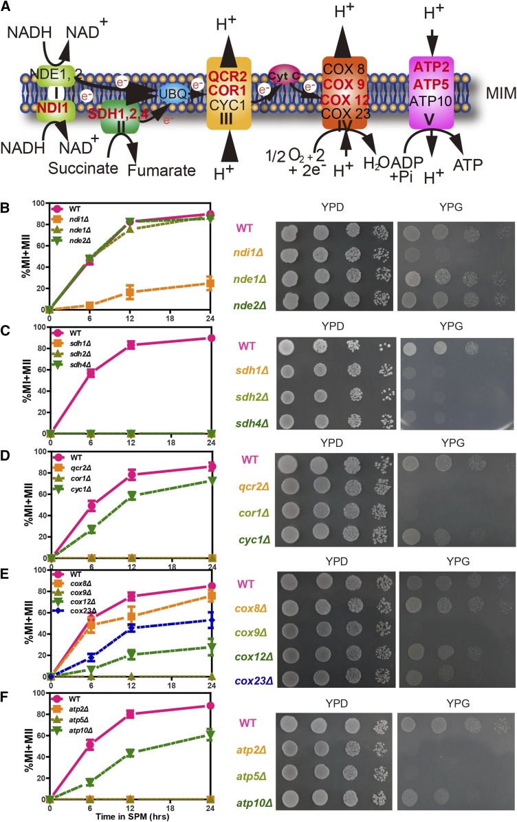
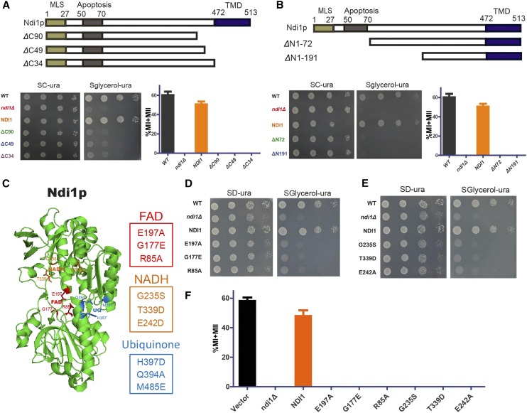
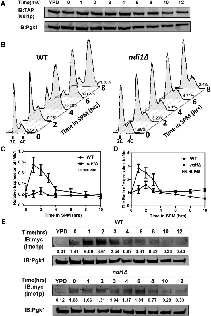
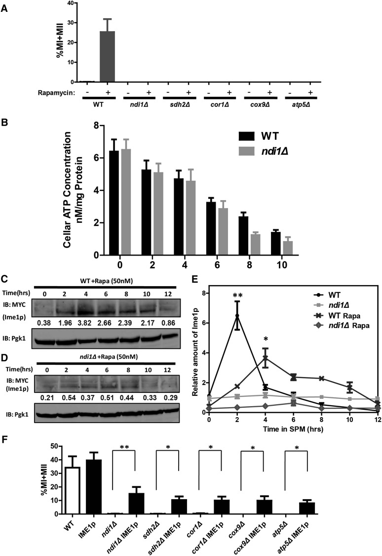
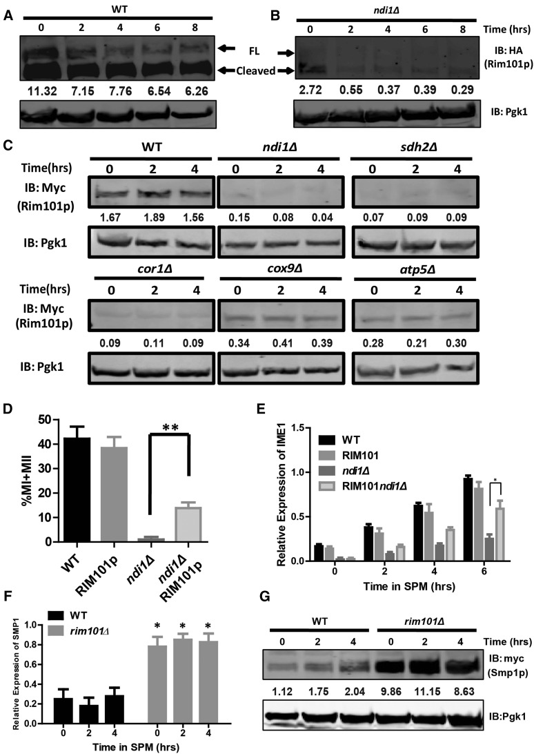
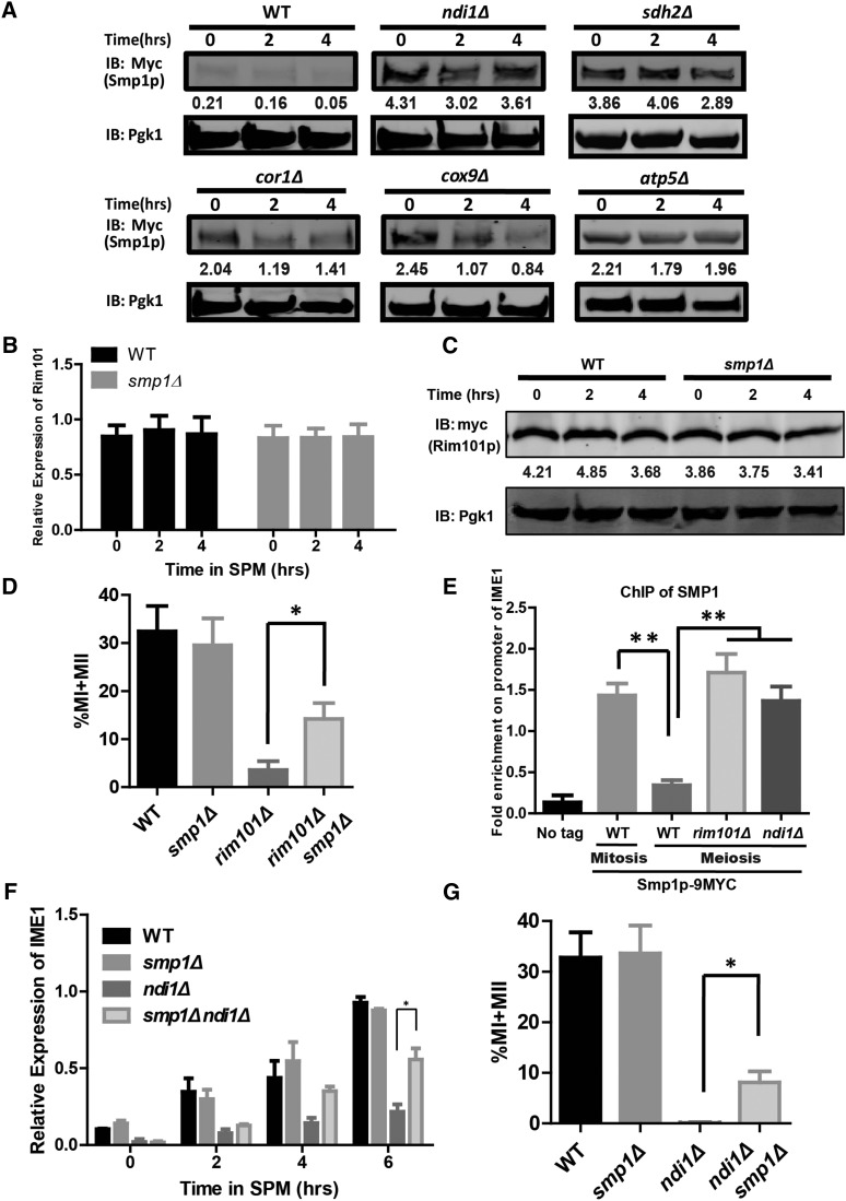
Meiosis is a specific type of cell division that is essential for sexual reproduction in most eukaryotes. Mitochondria are crucial cellular organelles that play important roles in reproduction, though the detailed mechanism by which the mitochondrial respiratory chain functions during meiosis remains elusive. Here, we show that components of the respiratory chain (Complexes I-V) play essential roles in meiosis initiation during the sporulation of budding yeast, Saccharomyces cerevisiae Any functional defects in the Complex I component Ndi1p resulted in the abolishment of sporulation. Further studies revealed that respiratory deficiency resulted in the failure of premeiotic DNA replication due to insufficient IME1 expression. In addition, respiration promoted the expression of RIM101, whose product inhibits Smp1p, a negative transcriptional regulator of IME1, to promote meiosis initiation. In summary, our studies unveiled the close relationship between mitochondria and sporulation, and uncover a novel meiosis initiation pathway that is regulated by the respiratory chain.

Keywords: NDI1; SMP1; meiosis initiation; respiratory chain; sporulation.


Figures

Figure 1
Figure 1
The ETC is essential for sporulation and respiration. (A) Schematic representation of Complexes I–V in the ETC. (B–F) Effects on sporulation and respiration of the deletion of key enzymes from Complexes I–V. Left panel: WT and mutant strains were induced to sporulate by transfer to SPM and analyzed at different time points. The divided nucleates, representing the sporulation efficiency, were stained with DAPI, visualized and counted under a microscope. Error bars indicate ± SEM (n = 3). Right panel: Yeast cells were spotted onto corresponding positions on YPD or YPG (a nonfermentable carbon source medium) plates by serial 10-fold dilutions (n = 4). WT: LW0066; ndi1Δ: LW0323; nde1Δ: LW0326; nde2Δ: LW0329; sdh1Δ: LW1327; sdh2Δ: LW1330; sdh4Δ: LW1333; qcr2Δ: LW1336; cor1Δ: LW1339; cyc1Δ: LW1342; cox8Δ: LW1345; cox9Δ: LW1348; cox12Δ: LW1351; cox23Δ: LW1354; atp2Δ: LW1357; atp5Δ: LW1360; atp10Δ: LW1363.
Figure 2
Figure 2
Effects of Ndi1p on sporulation and respiration. (A and B, top panel) Schematic representation of the domains of Ndi1p, including ΔC90 (Δ424–513 aa, LW1522), ΔC49 (Δ465–513 aa, LW1523), ΔC34 (Δ480–513 aa, LW1524), ΔN1–72 (Δ1–72 aa, LW1531), and ΔN1–191 (Δ1–191 aa, LW1532). MLS indicates the mitochondria localization signal; apoptosis indicates the apoptosis-related region; TMD indicates the transmembrane domain. (A and B, bottom panel) Effects of different Ndi1p truncations on respiration and sporulation. Left panel, yeast cells were spotted onto corresponding positions on SD-ura or SG-ura plates by serial 10-fold dilutions (n = 4). Right panel shows that the indicated strains were induced to sporulate by transfer to SPM, and the percentages of MI and MII cells were measured after 24 hr. Error bars indicate ± SEM (n = 3). (C) Schematic representation of key residues of the functional sites in Ndi1p, including the FAD-binding pocket (R85, G177, and E197), NADH-binding pocket (G235, E242, and T339), and ubiquinone (UQ)-binding site (Q394, H397, and M485). (D) Interaction between Ndi1p and FAD is essential for respiration. Yeast cells (WT: LW1399; ndi1Δ with an empty vector: LW1398; Ndi1p: LW1521; G177E: LW1534; and E197A: LW1533; R85A: LW1535) were established as described in A. (E) The NADH-binding pocket of Ndi1p is essential for respiration. Yeast cells (WT; ndi1Δ with an empty vector; Ndi1p; G235S: LW1536; T339D: LW1537 and E242A: LW1538) were established as described in A. (F) The FAD- and NADH-binding pockets of Ndi1p are essential for sporulation. The yeast cells shown in F were established as described in A. Error bars indicate ± SEM (n = 3).
Figure 3
Figure 3
DNA replication and Ime1p induction defects in ndi1Δ cells. (A) Expression of Ndi1p during sporulation. The WT strain expressing the Ndi1p-TAP allele (LW1542) was incubated in SPM, and samples were collected at different times after sporulation induction. Expression of Ndi1p-TAP was analyzed over time by immunoblotting using an anti-TAP antibody. Pgk1p served as a loading control. (B) Premeiotic DNA replication was inhibited in the ndi1Δ strain during sporulation. WT (LW0066) or ndi1Δ strains (LW0323) were incubated in SPM, and samples were collected at different times after induction. The DNA content was analyzed by flow cytometry to detect premeiotic DNA replication (2C–4C transition). The percentage of cells with a 4C DNA content was shown on the right of the corresponding 4C DNA peak. (n = 3). (C) Quantitative PCR analysis of the IME1 expression level in WT (LW0066) and NDI1 deletion strains (LW0323). Cells were harvested at the indicated time points. Total RNA was isolated and reverse transcribed, and IME1 mRNA levels were measured by quantitative PCR. The signals were normalized to the levels of NUP48. HK, Housekeeping gene. Error bars indicate ± SEM (n = 3). (D) Quantification of IME1 by Q-PCR relative to the 0 hr time point shown in (C). (E) Expression of Ime1p was detected in WT and ndi1Δ strains by immunoblotting during sporulation. WT (LW1543) or ndi1Δ (LW1544) cells expressing the IME1-3×Myc allele were incubated in SPM, and samples were collected at different times. Pgk1p served as a loading control. Quantification of Ime1p via western blotting normalized by Pgk1p was shown below the corresponding bands.
Figure 4
Figure 4
Respiration regulates meiosis initiation via Ime1p. (A) ETC-deficient cells could not be induced to sporulate by rapamycin in YPD medium. Indicated cells were grown to saturation in YPD for 24 hr at 30° and then treated with methanol (control) or rapamycin. The ratio of divided nuclei in cells was analyzed at 24 hr after induction using DAPI staining. Error bars indicate ± SEM (n = 3). WT: LW0066; ndi1Δ: LW0323; sdh2Δ: LW1330; cor1Δ: LW1339; cox9Δ: LW1348; atp5Δ: LW1360. (B) The intracellular ATP level of WT (LW0066) and ndi1Δ strains (LW0323) during rapamycin-induced sporulation. The cells were harvested at indicated time points and lysised by ATP determination lysis solution. The supernatants were processed and intracellular ATP concentrations were determined as described in the protocol of the ATP Assay Kit. Error bars indicate ± SEM (n = 5). (C and D) Expression of Ime1p was detected in WT and ndi1Δ strains by immunoblotting during rapamycin-induced sporulation. WT (LW1543) or ndi1Δ (LW1544) expressing the IME1-3×Myc allele were incubated in YPD and induced to sporulate with rapamycin; samples were collected at different times. Pgk1p served as a loading control. The relative amount of Ime1p was normalized with Pgk1p. (E) Quantitative results of Figure 4, C and D. * P < 0.05. (F) Overexpression of Ime1p rescued the sporulation defect in ETC-deficient strains (ndi1Δ, sdh2Δ, cor1Δ, cox9Δ, and atp5Δ). An Ime1p expression vector under control of the CUP1 promoter was generated and transformed into ETC-deficient strains. Cells were induced to sporulate as described in A and stained with DAPI after 24 hr. Error bars indicate ± SEM (n = 3). * P < 0.05 or ** P < 0.01. WT, ndi1Δ, sdh2Δ, cor1Δ, cox9Δ, and atp5Δ strains were the same as those shown in A; IME1: LW1563; ndi1ΔIME1: LW1564; sdh2ΔIME1: LW1565; cor1ΔIME1: LW1566; cox9ΔIME1: LW1567; atp5ΔIME1: LW1568.
Figure 5
Figure 5
Rim101p participates in meiosis initiation. (A and B) Expression and activation of Rim101p were detected in WT (LW1571) and ndi1Δ strains (LW1572) by immunoblotting during rapamycin-induced sporulation. FL, full-length Rim101p; Cleaved, cleaved Rim101p. Pgk1p was used as a loading control. The relative amount of Rim101p was normalized with Pgk1p and is shown below the corresponding band. (C) Expression of Rim101p was lower in ETC-deficient strains (ndi1Δ, sdh2Δ, cor1Δ, cox9Δ, and atp5Δ) than that in WT during meiosis. Pgk1p served as a loading control. The relative amount of Rim101p was normalized with Pgk1p and is shown below the corresponding band. WT: LW1545; ndi1Δ: LW1546; sdh2Δ: LW1547; cor1Δ: LW1548; cox9Δ: LW1549; atp5Δ: LW1550. (D) Overexpression of Rim101p rescued the sporulation defect in the NDI1 deletion strain. RIM101 was expressed in ndi1Δ cells, which were then induced to sporulate as described in A; the cells were stained with DAPI after 24 hr. Error bars indicate ± SEM (n = 3). ** P < 0.01. WT: LW0066; RIM101: LW1551; ndi1Δ: LW1552; ndi1ΔRIM101: LW1553. (E) Overexpression of Rim101p increased the transcription level of IME1 in ndi1Δ cells. The cells and procedures used to induce sporulation were the same as those described in D. The IME1 expression level in WT and NDI1 deletion strains was analyzed by real-time PCR. The signals were normalized to ACT1 levels. Error bars indicate ± SEM (n = 3). * P < 0.05. (F) Real-time PCR analysis of the SMP1 expression level in WT (LW0066) and RIM101 deletion strains (LW1560). The procedure was the same as that described in E. Error bars indicate ± SEM (n = 3). * P < 0.05. (G) The protein level of Smp1p was higher in rim101Δ cells (LW1573) than in WT cells (LW1366) during meiosis. Cells expressing the Smp1p-9×Myc allele were induced to sporulate with rapamycin, and samples were collected at different times. Pgk1p served as a loading control. The relative amount of Smp1p was normalized with Pgk1p and is shown below the corresponding band.
Figure 6
Figure 6
The IME1 repressor Smp1p is down-regulated by Rim101p during meiosis. (A) Repression of Smp1p was impaired in ETC-deficient strains (ndi1Δ, sdh2Δ, cor1Δ, cox9Δ, and atp5Δ) during meiosis. Cells expressing Smp1p-9×Myc were induced to sporulate with rapamycin, and samples were collected at different times. Pgk1p served as a loading control. The relative amount of Smp1p was normalized with Pgk1p and is shown below the corresponding band. WT: LW1366; ndi1Δ: LW1554; sdh2Δ: LW1555; cor1Δ: LW1556; cox9Δ: LW1557; atp5Δ: LW1558. (B) Real-time PCR analysis of the RIM101 expression level in WT (LW0066) and SMP1 deletion (LW1559) strains. The procedure was the same as that described in Figure 5F. Error bars indicate ± SEM (n = 3). (C) The protein level of Rim101p did not differ markedly between smp1Δ cells (LW1574) and WT cells (LW1545) during meiosis. The procedure was the same as that described in Figure 5G. The relative amount of Rim101p was normalized with Pgk1p and is shown below the corresponding band. (D) SMP1 deletion rescued the sporulation defect in rim101Δ cells. The procedure used to induce sporulation was the same as that described in Figure 5A, and cells were stained with DAPI after 24 hr. Error bars indicate ± SEM (n = 3). * P < 0.05. WT: LW0066; smp1Δ: LW1559; rim101Δ: LW1560; smp1Δrim101Δ: LW1561. (E) The different binding activities of Smp1p to the IME1 promoter in WT (LW1366), rim101Δ (LW1573), and ndi1Δ (LW1554) cells. Mitotic cells (in YPD medium) and meiotic cells (in rapamycin + YPD medium) were cross-linked with formaldehyde, and Smp1p was immunoprecipitated from chromatin extracts. The recovered DNA was quantified by real-time PCR using primers (IME1-P3-F/R) corresponding to the IME1 promoter. The signals were normalized to whole-cell DNA. Error bars indicate ± SEM (n = 3). * P < 0.05. (F) Deletion of SMP1 increased the transcription level of IME1 in ndi1Δ cells. The IME1 expression level in WT and NDI1 deletion strains was analyzed by real-time PCR. The cells and procedures used to induce sporulation were the same as those described in Figure 4A. Error bars indicate ± SEM (n = 3). * P < 0.05. (G) SMP1 deletion rescued the sporulation defect in ndi1Δ cells. Cells were induced to sporulate as described in Figure 4A and stained with DAPI after 24 hr. Error bars indicate ± SEM (n = 3). * P < 0.05. WT: LW0066; smp1Δ: LW1559; ndi1Δ: LW0323; smp1Δndi1Δ: LW1562.
Figure 7
Figure 7
Proposed model for the functional role of respiration during meiosis initiation. In addition to providing energy, respiration is involved in meiosis initiation during sporulation in yeast. Respiration stimulates expression of RIM101, followed by suppression of SMP1. Abolishment of Smp1p binding to the IME1 promoter facilitates IME1 transcription. The high expression level of IME1 initiates premeiotic DNA replication, ultimately initiating the sporulation process. The gray areas indicate known results from previous studies. The colored areas indicate the new findings from the current study.

Similar articles

Cited by

References

    1. Amaral A., Lourenco B., Marques M., Ramalho-Santos J., 2013. Mitochondria functionality and sperm quality. Reproduction 146: R163–R174. - PubMed
    1. Baek Y. U., Martin S. J., Davis D. A., 2006. Evidence for novel pH-dependent regulation of Candida albicans Rim101, a direct transcriptional repressor of the cell wall beta-glycosidase Phr2. Eukaryot. Cell 5: 1550–1559. - PMC - PubMed
    1. Benjamin K. R., Zhang C., Shokat K. M., Herskowitz I., 2003. Control of landmark events in meiosis by the CDK Cdc28 and the meiosis-specific kinase Ime2. Genes Dev. 17: 1524–1539. - PMC - PubMed
    1. Chacinska A., Boguta M., 2000. Coupling of mitochondrial translation with the formation of respiratory complexes in yeast mitochondria. Acta Biochim. Pol. 47: 973–991. - PubMed
    1. Church C., Goehring B., Forsha D., Wazny P., Poyton R. O., 2005. A role for Pet100p in the assembly of yeast cytochrome c oxidase: interaction with a subassembly that accumulates in a pet100 mutant. J. Biol. Chem. 280: 1854–1863. - PubMed

Publication types

MeSH terms

Substances