. 2018 Mar;208(3):1165-1179.
doi: 10.1534/genetics.117.300447. Epub 2018 Jan 3.

Deletion of a Long-Range Dlx5 Enhancer Disrupts Inner Ear Development in Mice

Affiliations

Deletion of a Long-Range Dlx5 Enhancer Disrupts Inner Ear Development in Mice

Kenneth R Johnson et al. Genetics. 2018 Mar.

Abstract

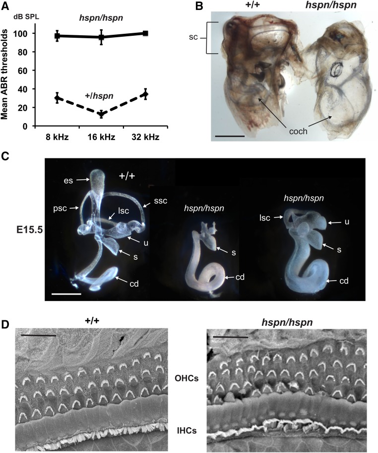
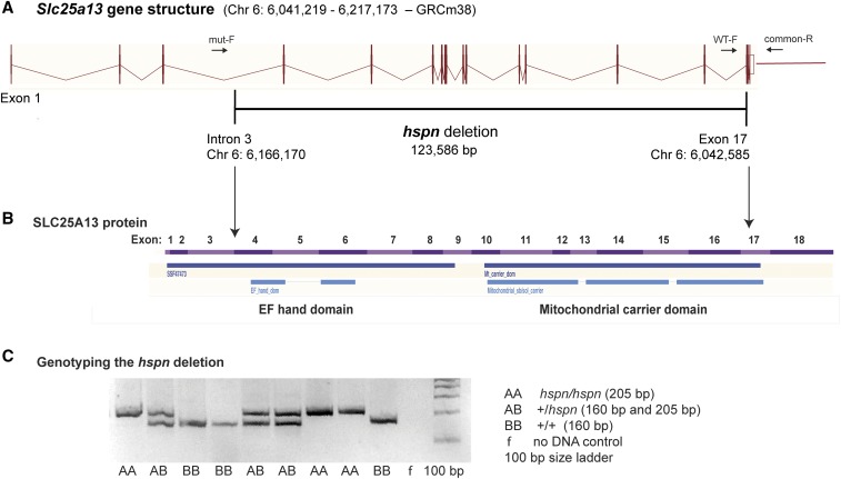
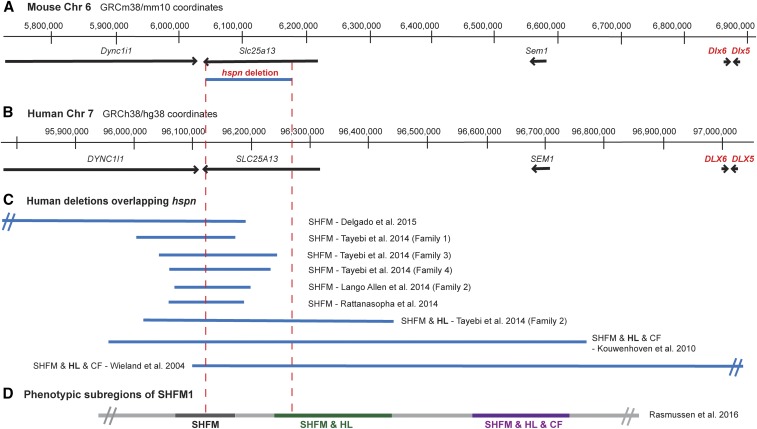
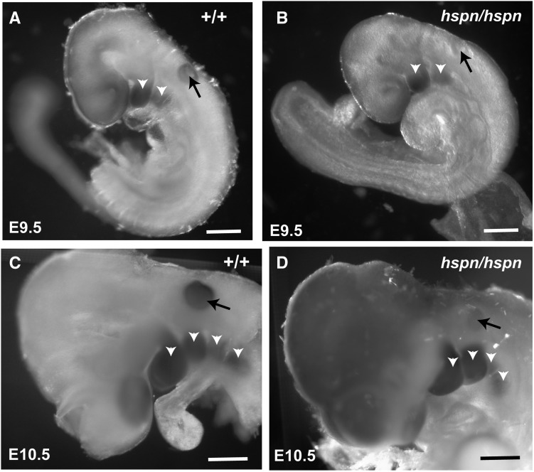
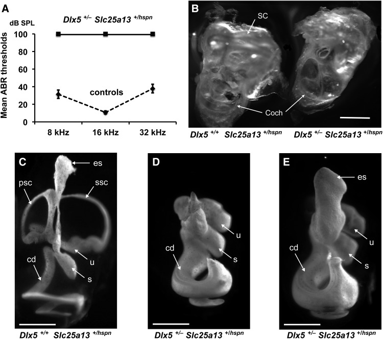
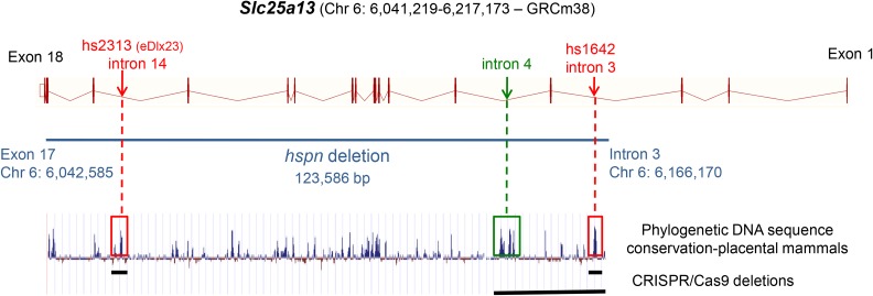
Distal enhancers are thought to play important roles in the spatiotemporal regulation of gene expression during embryonic development, but few predicted enhancer elements have been shown to affect transcription of their endogenous genes or to alter phenotypes when disrupted. Here, we demonstrate that a 123.6-kb deletion within the mouse Slc25a13 gene is associated with reduced transcription of Dlx5, a gene located 660 kb away. Mice homozygous for the Slc25a13 deletion mutation [named hyperspin (hspn)] have malformed inner ears and are deaf with balance defects, whereas previously reported Slc25a13 knockout mice showed no phenotypic abnormalities. Inner ears of Slc25a13hspn/hspn mice have malformations similar to those of Dlx5-/- embryos, and Dlx5 expression is severely reduced in the otocyst but not the branchial arches of Slc25a13hspn/hspn embryos, indicating that the Slc25a13hspn deletion affects otic-specific enhancers of Dlx5 In addition, transheterozygous Slc25a13+/hspn Dlx5+/- mice exhibit noncomplementation with inner ear dysmorphologies similar to those of Slc25a13hspn/hspn and Dlx5-/-embryos, verifying a cis-acting effect of the Slc25a13hspn deletion on Dlx5 expression. CRISPR/Cas9-mediated deletions of putative enhancer elements located within the Slc25a13hspn deleted region failed to phenocopy the defects of Slc25a13hspn/hspn mice, suggesting the possibility of multiple enhancers with redundant functions. Our findings in mice suggest that analogous enhancer elements in the human SLC25A13 gene may regulate DLX5 expression and underlie the hearing loss that is associated with split-hand/-foot malformation 1 syndrome. Slc25a13hspn/hspn mice provide a new animal model for studying long-range enhancer effects on Dlx5 expression in the developing inner ear.

Keywords: Dlx5; SHFM1; Slc25a13; distal enhancer; inner ear.


Figures

Figure 1
Figure 1
Inner ear phenotype of hspn/hspn mutant mice. (A) Hearing assessment by ABR. Average ABR thresholds of +/hspn heterozygous control mice (N = 32) tested at 5–12 weeks of age compared with those of hspn/hspn mutants (N = 7) tested at 4–5 weeks of age. The +/hspn control mice exhibited normal thresholds at all test frequencies (8, 16, and 32 kHz), whereas the highly elevated ABR thresholds of hspn/hspn mutant mice indicated profound hearing impairment. Error bars represent SD. (B) Cleared whole mounts of inner ears from 4-week-old adult mice. Inner ears of the adult hspn/hspn mutant shows reduced development of the semicircular canals (sc) and a much wider and shorter cochlea (coch) than that of the +/+ control. Bar, 1 mm. (C) Paintfills of the membranous labyrinths of inner ears from E15.5 embryos. Compared with the normal morphology of +/+ controls, inner ears of hspn/hspn mutants lack an endolymphatic sac (es) and duct, have a severe reduction in semicircular canal development, and exhibit a swollen and shortened cochlear duct (cd). The saccule (s) and cochlear duct were present in all hspn/hspn mutant inner ears examined; however, the extent of dorsal structure development varied. Posterior semicircular canals (psc) and superior semicircular canals (ssc) never developed in mutant inner ears, but there was variable development of the lateral semicircular canal (lsc) and utricle (u). Bar, 0.5 mm. (D) SEM surface images of the organ of Corti in the midapical region of the cochlea from a 4-week-old hspn/hspn mutant mouse and an age-matched +/+ control. Although a single row of inner hair cells (IHCs) and three rows of outer hair cells (OHCs) are present in both mutant and control mice, the hair cells of hspn/hspn mutant mice are disorganized, with an extra row of OHCs near the cochlear apex. Bar, 20 μm.
Figure 2
Figure 2
Molecular characterization of the Slc25a13hspn mutation. (A) Diagramatic representation of the exon–intron genomic structure of Slc25a13. The position of the 123,586-bp hspn deletion, extending from intron 3 to exon 17, is shown as a horizontal black line. Positions of PCR primers (mut-F, WT-F, and common-R) used to genotype mice for presence or absence of the hspn deletion are shown as ↔. The wild-type allele is amplified with the WT-F forward primer and the common-R reverse primer, and the hspn deletion allele is amplified with mut-F and common-R primers. (B) Structural diagram of the SLC25A13 protein. Regions of the protein encoded by the 18 exons of the Slc25a13 gene are shown as alternating pink and purple bands. Most of the protein is deleted by the hspn mutation (indicated by the area between the ↓’s), including both the EF-hand and mitochondrial domains. The structural diagrams for the Slc25a13 gene and SLC25A13 protein were derived from the Ensembl genome browser. (C) Results from the three-primer PCR assay for the presence or absence of the hspn deletion using the mut-F, WT-F, and common-R primers shown in (A). The PCR product size for the Slc25a13 allele with the hspn deletion is 205 bp, and the PCR product size for the wild-type (+) allele is 160 bp; both product sizes are detected in +/hspn heterozygotes.
Figure 3
Figure 3
Chr 6 map position of the mouse Slc25a13hspn deletion relative to Dlx5/6 and the corresponding SHFM1 region of human chromosome 7. (A) The region of mouse Chr 6 that corresponds to the human chromosome 7q21.3 region of SHFM1. Positions of the Dync1i1, Slc25a13, Shfm1, Dlx6, and Dlx5 genes are shown as horizontal black lines with arrowheads indicating the direction of transcription. The position of the Slc25a13hspn intragenic deletion is shown as a horizontal blue line. (B) The corresponding SHFM1 locus of human chromosome 7. Positions of the DYNCLI1, SLC25A13, SEM1, DLX6, and DLX5 genes are shown as horizontal black lines with arrowheads indicating the direction of transcription. (C) Deletions in SHFM1 patients that overlap with the position of the mouse Slc25a13hspn deletion (demarcated by the area between the vertical red dotted lines). The SHFM1 deletions (Wieland et al. 2004; Kouwenhoven et al. 2010; Lango Allen et al. 2014; Rattanasopha et al. 2014; Tayebi et al. 2014; Delgado and Velinov 2015) were associated with isolated SHFM, SHFM with hearing loss (SHFM & HL), or SHFM with hearing loss and craniofacial abnormalities (SHFM1 & HL & CF). The diverse SHFM1 phenotypes are thought to be the result of disrupted long-range enhancer effects on DLX5/DLX6 expression. (D) The three phenotypic subregions of the SHFM1 locus inferred from their correlations with chromosomal deletions and inversion break points (Rasmussen et al. 2016). The Slc25a13hspn deletion overlaps with part of the SHFM & HL subregion.
Figure 4
Figure 4
Decreased Dlx5 expression in the otic vesicle of Slc25a13hspn/hspn embryos. Whole mounts of embryos were hybridized with a Dlx5 antisense RNA probe. Wild-type Slc25a13 (+/+) embryos at E9.5 (A) and E10.5 (C) showed expression of Dlx5 in the branchial arches (open arrowheads) and in the otic vesicle (solid arrows). Although Slc25a13hspn/hspn (hspn/hspn) mutant embryos (B and D) showed strong Dlx5 expression in the branchial arches, expression appeared absent in the otic vesicle at E9.5 (B) and severely reduced in the otic vesicle at E10.5 (D). Bars, 500 μm.
Figure 5
Figure 5
The phenotype of Dlx5+/ Slc25a13+/hspn transheterozygotes is similar to that of Dlx5/ knockout mice. (A) Mice between 5 and 10 weeks of age were tested for ABR thresholds at 8-, 16-, and 32-kHz frequencies. All three Dlx5+/ Slc25a13+/hspn transheterozygotes lacked ABRs even at the highest test stimulus (100-dB SPL), whereas all eight control mice (three Dlx5+/+ Slc25a13+/+, two Dlx5+/+ Slc25a13+/hspn, and three Dlx5+/ Slc25a13+/+) exhibited normal thresholds. Error bars represent SD. (B) Cleared inner ears from an adult (10 weeks old) Dlx5+/+ Slc25a13+/hspn control and a Dlx5+/ Slc25a13+/hspn transheterozygous littermate. Inner ears of Dlx5+/ Slc25a13+/hspn transheterozygotes lack fully developed semicircular canals (SC) and have a malformed cochlea (Coch) with enlarged duct. Bar, 1 mm. (C–E) Paintfills of the membranous labyrinths of inner ears from E15.5 embryos. Inner ears of Dlx5+/+ Slc25a13+/hspn controls (C) have normal morphology, whereas inner ears of Dlx5+/ Slc25a13+/hspn transheterozygotes (D and E) lack dorsal structures and have malformed and thickened cochlear ducts. An enlarged endolymphatic sac was seen in one (E) but not all (D) inner ears of transheterozygotes. cd, cochlear duct; es, endolymphatic sac; psc, posterior semicircular canal; s, saccule; ssc, superior semicircular canal; u, utricle. Bars, 0.5 mm.
Figure 6
Figure 6
Putative enhancer elements within the Slc25a13hspn deletion. Locations of two putative enhancer elements, hs2313 (Chr6: 6,058,554–6,059,028; GRCm38) and hs1642 (Chr 6: 6,163,229–6,163,674), that lie within the hspn-deleted region (shown as horizontal blue line) of Slc25a13 were obtained from the VISTA Enhancer Browser (http://enhancer-test.lbl.gov/frnt_page.shtml). The hs2313 element is equivalent to the eDlx#23 enhancer (Birnbaum et al. 2012). DNA sequences of both elements show a high degree of phylogenic conservation for noncoding DNA (peaks enclosed by red boxes). An additional noncoding region of high sequence conservation occurs in intron 4 (peak enclosed by green box). Specific deletions corresponding to these conserved regions were created using CRISPR/Cas9 genome editing, and the extent of these deletions are shown by horizontal black lines at the bottom of the figure. The gene structure diagram for Slc25a13 is derived from the Ensembl genome browser, and the phylogenetic conservation diagram is from the University of California, Santa Cruz Genome Browser comparative genomics track.

Similar articles

Cited by

References

    1. Acampora D., Merlo G. R., Paleari L., Zerega B., Postiglione M. P., et al. , 1999. Craniofacial, vestibular and bone defects in mice lacking the distal-less-related gene Dlx5. Development 126: 3795–3809. - PubMed
    1. Ahituv N., Zhu Y., Visel A., Holt A., Afzal V., et al. , 2007. Deletion of ultraconserved elements yields viable mice. PLoS Biol. 5: e234. - PMC - PubMed
    1. Bernardini L., Palka C., Ceccarini C., Capalbo A., Bottillo I., et al. , 2008. Complex rearrangement of chromosomes 7q21.13-q22.1 confirms the ectrodactyly-deafness locus and suggests new candidate genes. Am. J. Med. Genet. A. 146A: 238–244. - PubMed
    1. Birnbaum R. Y., Everman D. B., Murphy K. K., Gurrieri F., Schwartz C. E., et al. , 2012. Functional characterization of tissue-specific enhancers in the DLX5/6 locus. Hum. Mol. Genet. 21: 4930–4938. - PMC - PubMed
    1. Brown K. K., Reiss J. A., Crow K., Ferguson H. L., Kelly C., et al. , 2010. Deletion of an enhancer near DLX5 and DLX6 in a family with hearing loss, craniofacial defects, and an inv(7)(q21.3q35). Hum. Genet. 127: 19–31. - PMC - PubMed

Publication types

LinkOut - more resources