Review
. 2018 Jan;208(1):53-78.
doi: 10.1534/genetics.117.300082.

Invading, Leading and Navigating Cells in Caenorhabditis elegans: Insights into Cell Movement in Vivo

Affiliations
Review

Invading, Leading and Navigating Cells in Caenorhabditis elegans: Insights into Cell Movement in Vivo

David R Sherwood et al. Genetics. 2018 Jan.

Abstract

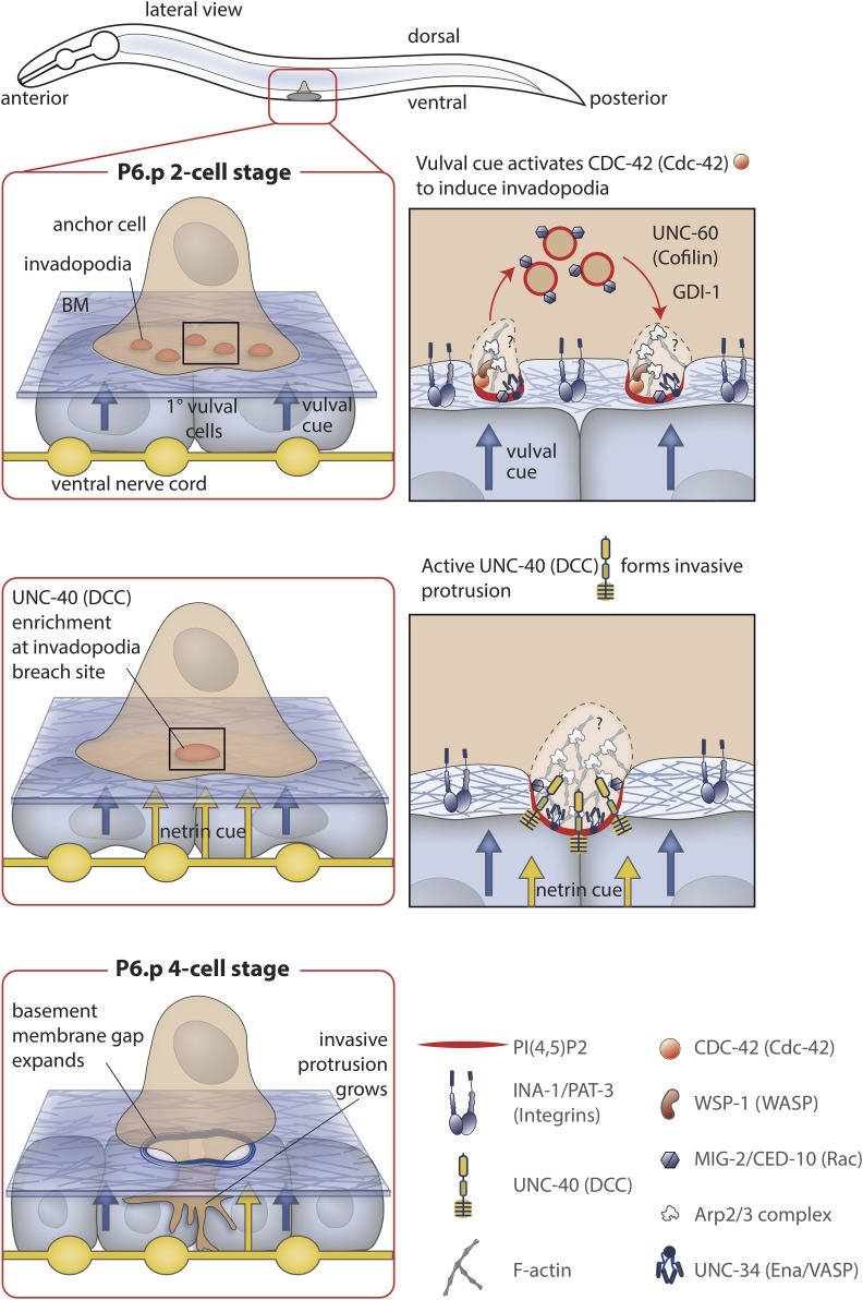
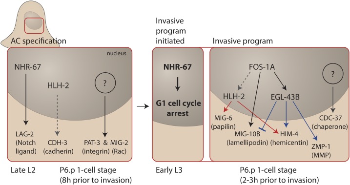
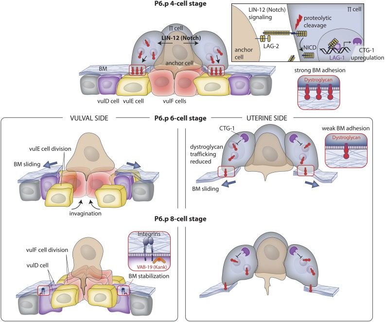
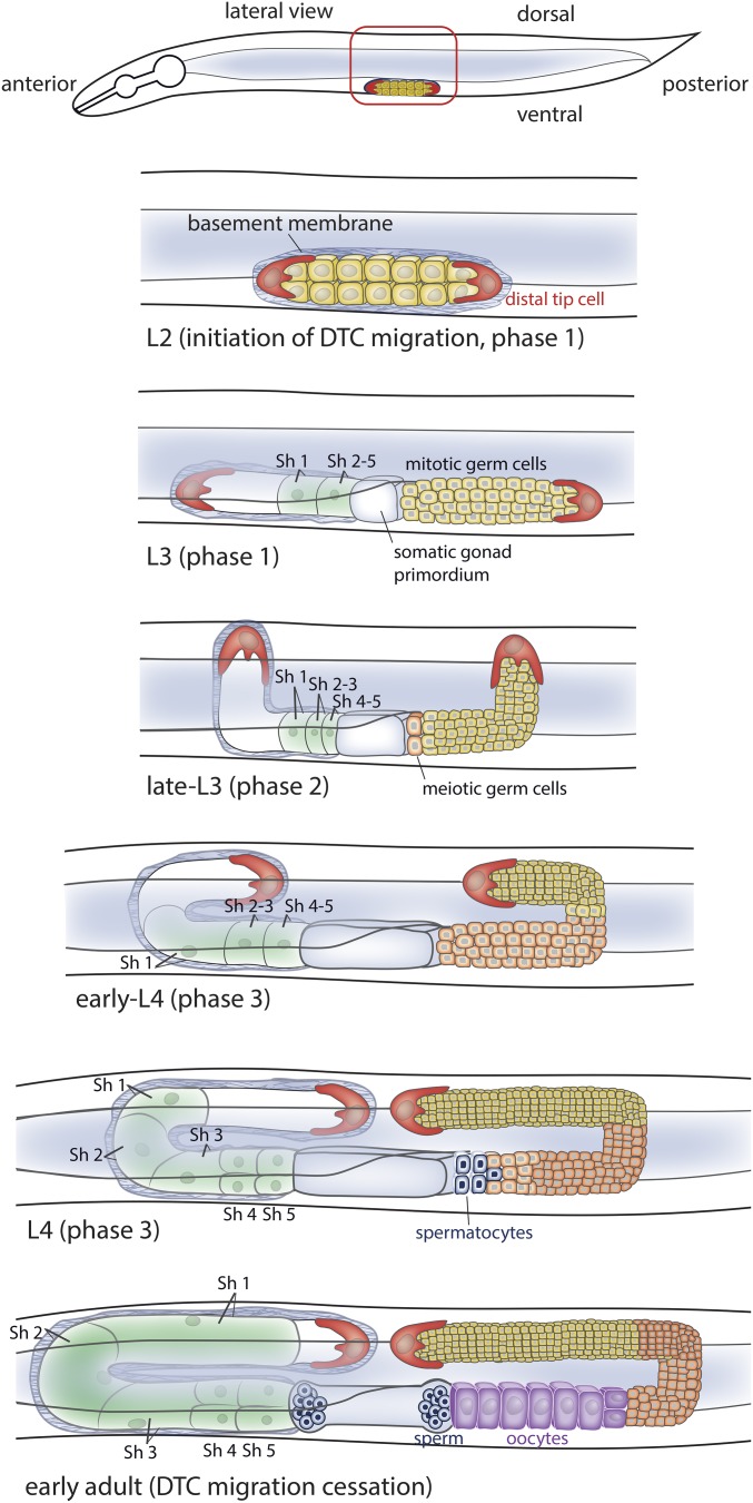
Highly regulated cell migration events are crucial during animal tissue formation and the trafficking of cells to sites of infection and injury. Misregulation of cell movement underlies numerous human diseases, including cancer. Although originally studied primarily in two-dimensional in vitro assays, most cell migrations in vivo occur in complex three-dimensional tissue environments that are difficult to recapitulate in cell culture or ex vivo Further, it is now known that cells can mobilize a diverse repertoire of migration modes and subcellular structures to move through and around tissues. This review provides an overview of three distinct cellular movement events in Caenorhabditis elegans-cell invasion through basement membrane, leader cell migration during organ formation, and individual cell migration around tissues-which together illustrate powerful experimental models of diverse modes of movement in vivo We discuss new insights into migration that are emerging from these in vivo studies and important future directions toward understanding the remarkable and assorted ways that cells move in animals.

Keywords: C. elegans; F-actin; FGF pathway; Wnt pathway; WormBook; basement membrane; cell invasion; cell migration; cell signaling; integrin; netrin pathway.


Figures

Figure 1
Figure 1
AC invasion, a BM invasion event. Prior to invasion, the AC is positioned over the epidermal P6.p cell, which it induces to a 1° vulval precursor cell fate. The 1° fated P6.p divides three times. Invasion occurs at the time of the division from the P6.p 2-cell stage to P6.p 4-cell stage. Top panel: Prior to breaching the BM, AC invadopodia form along the AC’s invasive cell membrane, depress the BM, and then disassemble. Approximately 10 invadopodia are present at any one time. Invadopodia formation is stimulated by a cue from the 1° vulval cells that activates the Rho GTPase CDC-42. CDC-42 activates WSP-1, which presumably stimulates F-actin production through actin polymerization nucleators such as the Arp2/3 complex. UNC-34 (Ena/VASP) may also contribute to F-actin formation. Invadopodia generation is also dependent on an invadopodial membrane rich in PI(4,5)P2 and containing the lipid-anchored Rac GTPases, CED-10 and MIG-2. The invadopodial membrane is recycled through the endolysosome, and its trafficking is dependent on UNC-60 (cofilin) and the Rab GDP dissociation inhibitor (GDI-1). The integrin heterodimer INA-1/PAT-3 is required for trafficking of all known invadopodial components to the plasma membrane. Middle panel: when an invadopodium breaches the BM, the netrin receptor UNC-40 (DCC) traffics to the breach site, and is activated by its ligand UNC-6 (netrin) secreted by the underlying ventral nerve cord. UNC-40 recruits the actin regulators UNC-34 (Ena/VASP) and the Rac GTPases, which shuts down further invadopodia formation. Lower Panel: UNC-40 (DCC) directs the formation of a large invasive protrusion that expands the opening in the BM by degrading and physically displacing BM.
Figure 2
Figure 2
Transcription factors that specify invasive cell fate. Left: The AC is specified during the late L2 stage by the action of several transcription factors, including NHR-67 (Tailless) and HLH-2 (Daughterless/E proteins) that drive the expression of genes encoding LAG-2 (Notch ligand) and CDH-3 (cadherin), respectively, as well as other unknown transcription factors that promote the expression of genes encoding PAT-3 (β-integrin) and MIG-2 (Rac). Right: During the early L3 stage NHR-67 directs the AC into G1 cell cycle arrest, which allows full invasive fate differentiation. A central transcription factor that operates following G1 arrest is FOS-1A (Fos), which promotes the expression of the transcription factors HLH-2 and EGL-43B (EVI1). These transcription factors regulate the expression of several invasion effector genes that encode MIG-6 (papilin), MIG-10B (lamelipodin), HIM-4 (hemicentin), and ZMP-1 (MMP). Other transcription factors remain to be discovered as genes encoding proteins such as CDC-37 (Hsp90 cochaperone) are expressed in the AC and promote invasion, but are not regulated by the FOS-1A transcriptional network.
Figure 3
Figure 3
BM sliding following AC invasion widens the breach. Top panel (P6.p 4-cell stage): during invasion, the AC activates LIN-12 (Notch) signaling in neighboring uterine π cells via the ligand LAG-2 (Delta; see upper right box). Notch activation leads to proteolytic release of the LIN-12 (Notch) intracellular domain (NICD), which enters the nucleus, associates with LAG-1 (CSL), and upregulates expression of the gene encoding CTG-1 (Sec14-GOLD protein). Middle panel left (P6.p 6-cell stage): vulval and uterine tissue growth and vulval cell invagination apply forces on the BM that drive its shifting. VulF cells begin to invaginate and the vulE cells divide, lose contact with the BM, and allow the BM to slide over these cells, thus widening the BM gap. Middle panel right: CTG-1 activity in the uterine π cells inhibits the trafficking of the BM adhesion receptor dystroglycan to the cell-BM interface, weakening BM adhesion, and allowing the BM to move on the uterine side of the BM. Lower panel left (P6.p 8-cell stage): the vulF cells divide and further invaginate with the vulE cells. The BM stops shifting over the nondividing vulD cell, which sets the width of the opening of the BM gap. Integrin and VAB-19 (KANK) localize to the vulD-BM interface to stabilize BM adhesion. Lower panel right: dystroglycan levels continue to be reduced at the interface, allowing the BM to slide to a position determined by the underlying vulD cell.
Figure 4
Figure 4
DTC migration, a leading cell that shapes an organ. The pair of DTCs (one on the anterior the other on the posterior arm of the basement membrane enwrapped gonad) initiate migration at the L2 larval stage. During the L2 and L3 larval stages (phase 1 of migration), the DTCs move ventrally away from each other along the BMs of the ventral body wall muscles (data not shown) toward the anterior (head) and posterior (tail) of the animal. In the rest of the figure, the posterior gonad arm (right side) shows the germ cells from the L3 stage onward, while the anterior arm (left side) shows the basement membrane and five pairs of sheath cells (sh1-5) that cover the germ cells. The sheath cells follow the path of the DTCs. The digestive tube is shown in light gray and the anterior gonad arm passes underneath it to the other side of the animal. During the late-L3 stage, both DTCs turn 90° and move from the ventral to dorsal surface (phase 2 of migration), moving along the BM of the lateral epidermis. At the early L4 stage, the DTCs turn 90° and move back to the midsection along the BM of dorsal body wall muscles during the L4 stage (phase 3 of migration). DTC migration ceases in the early adult.
Figure 5
Figure 5
DTC migration timing, BM interactions, polarization, and gene regulation. Only the posterior gonad arm is shown. (A) The timing of DTC migration shown is at 20°. (B–D) Details of proteins and interactions that regulate DTC migration are described in the text and outlined here for a global view. Note, the “Wnt ⊣” shown in (C) represents a hypothetical possible function for Wnt in inhibiting polarization along the anterior-posterior axis during phase 2 of DTC migration.
Figure 6
Figure 6
Examples of DTC migration defects. Only the posterior gonad arm is shown. (A) Wild type migration, (B) a gon-1 mutant where no DTC migration occurs, (C) an ndk-1 mutant where DTC migration is incomplete, (D) a src-1 mutant and an unc-5 mutant where DTC migration shows pathfinding defects, (E) reduction of cacn-1 by RNAi leads to extended DTC migration (a cessation of migration defect).
Figure 7
Figure 7
SM migration, a cell navigation event. Top panels: two SMs are specified by LIN-12 (Notch) signaling and the action of the zinc finger transcriptional regulator SEM-4 in the tail of the L1 larva and migrate independently of each other through the body of the worm over the course of the L2 and L3 larval stages until reaching the central gonad (shown with darker shading). The transcriptional regulator LIN-42 (Period) prevents the SMs from dividing precociously during their migration. Only one SM is shown for simplicity. Bottom panel: SMs are directed to the proper location by an EGL-17 (FGF) signal emanating from the central gonad region and vulval precursor cells, and by an additional unknown signal originating from nongonadal tissue. In addition to these attractive signals, an unidentified cue, also originating in the gonad, repels the SMs. Both EGL-17 (FGF)-dependent attractive and gonad-dependent repulsive signals are sensed by the FGF receptor EGL-15 in the SM, but different isoforms (5A and 5B) respond to attractive and repulsive cues. Signals are transduced to LET-60 (Ras) via the adaptor molecule SEM-5 (Grb2), which may also communicate to Arp2/3 complex-driven actin assembly. EGL-17 (FGF)-independent attraction is less well-understood, but depends on various molecules with potential roles in cell adhesion and motility such as UNC-53 (Nav2), UNC-71 (ADAM), and UNC-73 (Trio).

References

    1. Abella J. V., Galloni C., Pernier J., Barry D. J., Kjaer S., et al. , 2016. Isoform diversity in the Arp2/3 complex determines actin filament dynamics. Nat. Cell Biol. 18: 76–86. - PubMed
    1. Altincicek B., Fischer M., Luersen K., Boll M., Wenzel U., et al. , 2010. Role of matrix metalloproteinase ZMP-2 in pathogen resistance and development in Caenorhabditis elegans. Dev. Comp. Immunol. 34: 1160–1169. - PubMed
    1. Aman A., Piotrowski T., 2010. Cell migration during morphogenesis. Dev. Biol. 341: 20–33. - PubMed
    1. Andrew D. J., Ewald A. J., 2010. Morphogenesis of epithelial tubes: insights into tube formation, elongation, and elaboration. Dev. Biol. 341: 34–55. - PMC - PubMed
    1. Antebi A., Culotti J. G., Hedgecock E. M., 1998. daf-12 regulates developmental age and the dauer alternative in Caenorhabditis elegans. Development 125: 1191–1205. - PubMed

Publication types

LinkOut - more resources