. 2018 Jan 5;359(6371):48-55.
doi: 10.1126/science.aan1078.

Transferrin receptor 1 is a reticulocyte-specific receptor for Plasmodium vivax

Affiliations

Transferrin receptor 1 is a reticulocyte-specific receptor for Plasmodium vivax

Jakub Gruszczyk et al. Science. .

Abstract

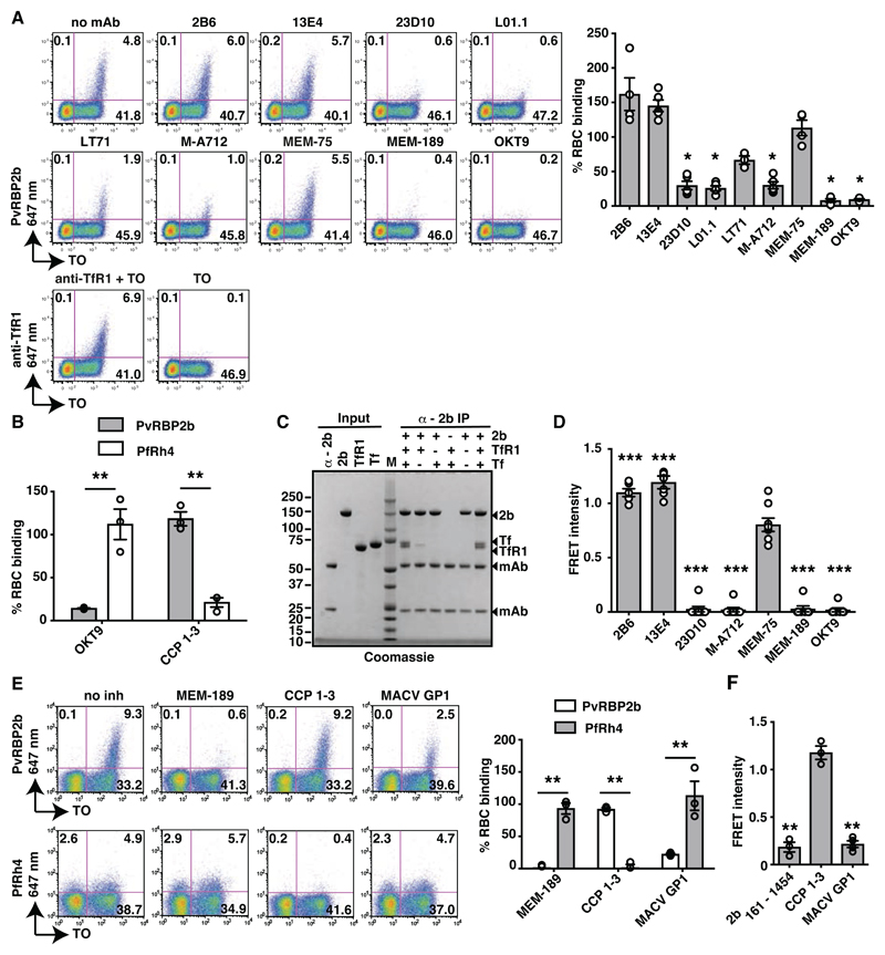
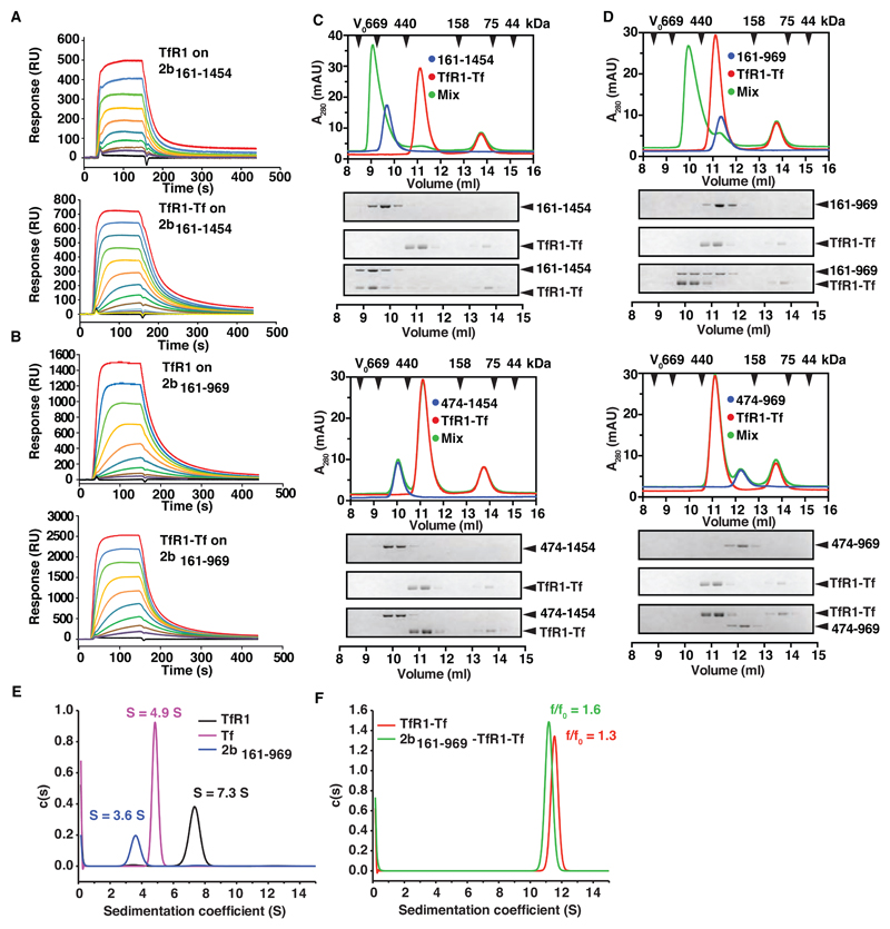
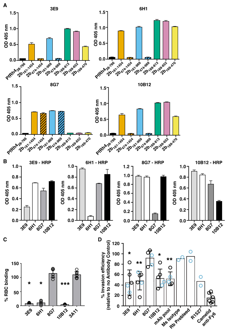
Plasmodium vivax shows a strict host tropism for reticulocytes. We identified transferrin receptor 1 (TfR1) as the receptor for P. vivax reticulocyte-binding protein 2b (PvRBP2b). We determined the structure of the N-terminal domain of PvRBP2b involved in red blood cell binding, elucidating the molecular basis for TfR1 recognition. We validated TfR1 as the biological target of PvRBP2b engagement by means of TfR1 expression knockdown analysis. TfR1 mutant cells deficient in PvRBP2b binding were refractory to invasion of P. vivax but not to invasion of P. falciparum Using Brazilian and Thai clinical isolates, we show that PvRBP2b monoclonal antibodies that inhibit reticulocyte binding also block P. vivax entry into reticulocytes. These data show that TfR1-PvRBP2b invasion pathway is critical for the recognition of reticulocytes during P. vivax invasion.


Figures

Fig. 1
Fig. 1. PvRBP2b binds TfR1 on the reticulocyte surface.
(A). PvRBP2b161-1454 binding in the presence of anti-TfR1 mAbs analysed by flow cytometry. Left: Dot plots of PvRBP2b161-1454 binding (y-axis) to reticulocytes stained with thiazole orange (TO, x-axis). Right: normalized binding results where PvRBP2b161-1454 binding in the absence of mAbs was arbitrarily assigned to be 100%. (B) PvRBP2b161-1454 and PfRh428-766 binding were evaluated by flow cytometry with the addition of anti-TfR1 mAb OKT9 or CCP 1-3. PvRBP2b161-1454 and PfRh4 binding in buffer were arbitrarily assigned to be 100%. (C) Eluates of individual or mixtures of proteins immuno-precipitated with anti-PvRBP2b mAb analyzed by SDS-PAGE. + or – indicates protein present or absent. Molecular weight marker (M). (D) Anti-TfR1 mAbs inhibit PvRBP2b-TfR1 complex formation in the FRET-based assay. The FRET signal was relative to “no mAb” control. (E) Binding of PvRBP2b161-1454 and PfRh428-766 in the presence of anti-TfR1 mAb MEM-189, CCP 1-3 and MACV GP1. Left: dot plots showing PvRBP2b161-1454 (top) and PfRh428-766 binding (bottom). Right: normalized binding results where PvRBP2b161-1454 and PfRh428-766 binding in the presence of buffer was arbitrarily assigned to be 100%. (F) MACV GP1 inhibits PvRBP2b161-1454-TfR1 complex formation monitored by FRET assay. For (A), (B), (D), (E) and (F), Mean ± S.E.M., n ≥3, open dots represent biological replicates. Mann-Whitley test was used for (A) and (D) where MEM-75 was considered non-inhibitory and t-tests was used for (B), (E) and (F), * P ≤ 0.05, ** P ≤ 0.001.
Fig. 2
Fig. 2. Crystal structure of the N-terminal domain of PvRBP2b and its functional requirement.
(A) Structure of the N-terminal domain of PvRBP2b from amino acid 169 to 470 shown in two orthogonal views. (B) Electrostatic surface potential on the PvRBP2b structure. (C) Superimposition of the PvRBP2b structure (green) with PvRBP2a (purple) and PfRh5 (orange). The PDB ID codes for PfRh5 and PvRBP2a are 4WAT and 4Z8N, respectively. (D) Crystal structure of the N-terminal domain superimposed with SAXS ab initio bead model of PvRBP2b169-470 (left) and PvRBP2b169-652 (right). (E) Sliding window analysis showing nucleotide diversity (π) values and Tajima’s D statistic in PvRBP2b. The grey box refers to a highly polymorphic region at amino acid positions 169 to 470 that appears to be under balancing selection. (F) Schematic representation of full-length PvRBP2b and recombinant protein fragments (left). Signal peptide (SP), transmembrane domain (TM) and N-terminal domain (yellow) are indicated. (G) PvRBP2b binding results by flow cytometry where PvRBP2b161-1454 binding was arbitrarily assigned to be 100%. (H) Unlabeled recombinant PvRBP2b fragments or PfRh4 were mixed at 10-fold molar excess relative to the labeled PvRBP2b161-1454-TfR1 FRET pair. The FRET intensity was relative to buffer control. For (G to H), Mean ± S.E.M, n = 4, open dots represent biological replicates. The Mann-Whitley test was used to calculate the P value using the binding of 2b474-1454 that was considered no binding, * P≤ 0.05, ** P ≤ 0.001.
Fig. 3
Fig. 3. PvRBP2b binds to TfR1-Tf to form a stable ternary complex.
PvRBP2b161-1454 (A) and PvRBP2b161-969 (B) were coupled covalently to a biosensor chip to probe binding of TfR1 (concentration range assayed: 2 µM to 7.5 nM, top panels) and TfR1-Tf complexes (concentration range of TfR1-Tf complexes assayed: 2 μM:4 μM to 1.8 nM:3.9 nM (A) and 2 μM:4 μM to 7.5 nM:15 nM (B), bottom panels). (C) and (D) Complex formation between PvRBP2b, TfR1 and Tf analyzed by analytical size exclusion chromatography (SEC). PvRBP2b-TfR1-Tf ternary complex can be observed for PvRBP2b161-1454 (C, top panel) and PvRBP2b161-969 (D, top panel). Two corresponding truncations of the N-terminal domain, PvRBP2b474-1454 (C, bottom panel) and PvRBP2b474-969 (D, bottom panel) do not interact with the TfR1-Tf binary complex. The exclusion volume (V0) of the columns and the elution volumes of selected marker proteins are indicated with black arrowheads. Lower part: Coomassie-Blue stained SDS-PAGE gels of the fractions obtained from SEC. (E-F) Continuous sedimentation coefficient distributions derived from fitting sedimentation velocity data to a c(s) sedimentation model. (E) c(s) distributions for TfR1 (black line), Tf (magenta line) and PvRBP2b161-969 (blue line). (F) c(s) distributions for the TfR1-Tf complex (red line), and PvRBP2b161-969-TfR1-Tf complex (green line).
Fig. 4
Fig. 4. Deletions in TFRC reduce TfR1 surface expression, abolish PvRBP2b binding and inhibit P. vivax invasion.
(A) Expression of TfR1, BSG and GYPA on the surface of jkRBCs, TfR1 mutants, ∆BSG null and cultured erythrocytes (cRBCs) as measured by flow cytometry. The right most panels show cytospin analysis of cells stained with May-Grünwald Giemsa staining technique. (B) TfR1∆G217 mutation in TfR1 abrogates PvRBP2b binding as observed using analytical SEC. (C) Quantitative surface proteomics demonstrate specific reduction in TfR1 protein levels in TfR1 mutants compared to wildtype jkRBCs. Levels of Tf, the binding partner for TfR1, are similarly reduced. Significance A was used to estimate p-values, and a minimum of 2 peptides were required for protein quantitation. (D) Binding of recombinant PvRBP2b fragments to jkRBCs, TfR1mutants, ∆BSG and cRBCs cells are shown in blue. Negative controls of unstained cells and isotype control stained cells are shown in the white and orange lines respectively. Compilation of results from PvRBP2b fragment binding to jkRBCs, TfR1 mutants, ∆BSG and cRBCs (right panel). Mean ± S.E.M, n = 3. (E) Comparison of invasion efficiency between jkRBCs and TfR1 mutant cell lines with either P. vivax or P. falciparum. The data shown are averages and the standard error of the mean from between four to five biological replicates shown as open dots. P value was calculated using a paired, two-tailed T test, **** P ≤ 0.0001 and ns is non-significant.
Fig. 5
Fig. 5. Anti-PvRBP2b mAbs inhibit reticulocyte binding and P. vivax invasion in Brazilian and Thai clinical isolates.
(A) ELISA plates were coated with equimolar concentrations of each recombinant fragment and detection with anti-PvRBP2b mAbs 3E9, 6H1, 8G7 and 10B12 are shown. (B) Competition ELISA using immobilized PvRBP2b incubated with un-conjugated anti-2b mAbs (x-axis) and detected with 3E9-HRP, 6H1-HRP, 8G7-HRP and 10B12-HRP as indicated. For (A) and (B) error bars represent range showing the variability of duplicate measures. (C) PvRBP2b161-1454 binding in the presence of anti-PvRBP2b mAbs 3E9, 6H1, 8G7 and 10B12 was analysed by flow cytometry. Normalized binding results where PvRBP2b161-1454 binding in the absence of mAbs was arbitrarily assigned to be 100%. The anti-PvRBP2a mAb 3A11 was used as a negative antibody control. Mean ± S.E.M, n = 5, open dots represent biological replicates. The Kruskal-Wallis test was used to calculate the P value using 8G7 binding as no inhibition, *P ≤ 0.05, ***P ≤ 0.0001. (D) Invasion of P. vivax in Brazilian (blue open dots) and Thai (black open dots) clinical isolates in the presence of anti-PvRBP2b 3E9, 6H1, 8G7 and 10B12, pooled mAbs (each mAb at one third of final concentration), mouse isotype control, purified rabbit prebleed IgG, purified total IgG of anti-PvRBP2b polyclonal antibody R1527 and camelid anti-Fy6 mAb. Antibodies were added in concentrations from 25 µg/ml to 125 µg/ml except for the camelid anti-Fy6 mAb which was used at 25 µg/ml. Mean ± SD, n from 2 to 6, open dots represent biological replicates. For experiments with n > 2, we used the Kolmogorov-Smirnov test to compare 8G7 to 3E9, 6H1 and 10B12, *P ≤ 0.05, **P ≤ 0.001.

Similar articles

Cited by

References

    1. Maier AG, et al. Plasmodium falciparum erythrocyte invasion through glycophorin C and selection for Gerbich negativity in human populations. Nat Med. 2003;9:87–92. - PMC - PubMed
    1. Crosnier C, et al. Basigin is a receptor essential for erythrocyte invasion by Plasmodium falciparum. Nature. 2011;480:534–537. - PMC - PubMed
    1. Sim BK, Chitnis CE, Wasniowska K, Hadley TJ, Miller LH. Receptor and ligand domains for invasion of erythrocytes by Plasmodium falciparum. Science. 1994;264:1941–1944. - PubMed
    1. Tham W-H, et al. Complement receptor 1 is the host erythrocyte receptor for Plasmodium falciparum PfRh4 invasion ligand. Proc Natl Acad Sci U S A. 2010;107:17327–17332. - PMC - PubMed
    1. Miller LH, Mason SJ, Dvorak JA, McGinniss MH, Rothman IK. Erythrocyte receptors for (Plasmodium knowlesi) malaria: Duffy blood group determinants. Science. 1975;189:561–563. - PubMed

Publication types

MeSH terms