Review
. 2018 Feb;14(2):94-105.
doi: 10.1038/nrneurol.2017.175. Epub 2018 Jan 5.

Adulthood leukodystrophies

Affiliations
Review

Adulthood leukodystrophies

Wolfgang Köhler et al. Nat Rev Neurol. 2018 Feb.

Abstract

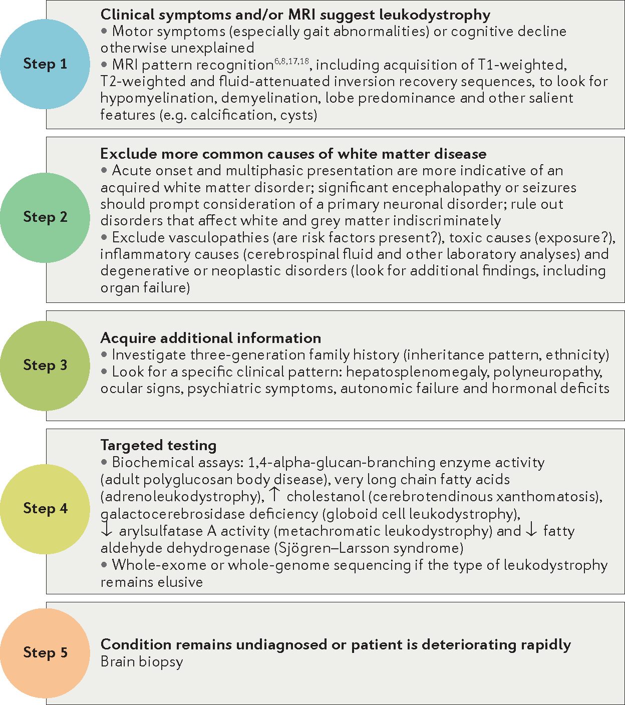
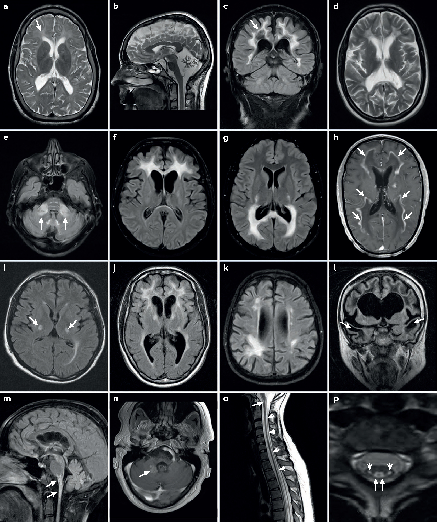
The leukodystrophies are a group of inherited white matter disorders with a heterogeneous genetic background, considerable phenotypic variability and disease onset at all ages. This Review focuses on leukodystrophies with major prevalence or primary onset in adulthood. We summarize 20 leukodystrophies with adult presentations, providing information on the underlying genetic mutations and on biochemical assays that aid diagnosis, where available. Definitions, clinical characteristics, age of onset, MRI findings and treatment options are all described, providing a comprehensive overview of the current knowledge of the various adulthood leukodystrophies. We highlight the distinction between adult-onset leukodystrophies and other inherited disorders with white matter involvement, and we propose a diagnostic pathway for timely recognition of adulthood leukodystrophies in a routine clinical setting. In addition, we provide detailed clinical information on selected adult-onset leukodystrophies, including X-linked adrenoleukodystrophy, metachromatic leukodystrophy, cerebrotendinous xanthomatosis, hereditary diffuse leukoencephalopathy with axonal spheroids, autosomal dominant adult-onset demyelinating leukodystrophy, adult polyglucosan body disease, and leukoencephalopathy with vanishing white matter. Ultimately, this Review aims to provide helpful suggestions to identify treatable adulthood leukodystrophies at an early stage in the disease course.


Conflict of interest statement

Competing interests statement

The authors declare no competing interests.

Figures

Figure 1 |
Figure 1 |. Diagnostic pathway for adulthood leukodystrophies.
Figure 2 |
Figure 2 |. Characteristic MRI features of adult leukodystrophies.
a | Hypomyelination in an adult with a Pol-III-related leukodystrophy. Arrow indicates moderately increased T2-weighted signal in affected white matter. Note relative atrophy, which is common in adults with hypomyelination. b | Severe cerebellar atrophy in a 24-year-old female with Pol-III-related hypomyelinating leukodystrophy. c | Diffuse cerebral white matter changes in a 61-year-old male with vanishing white matter disease. Note rarefaction of affected white matter (arrow) on fluid-attenuated inversion recovery images. d | Periventricular predominance of diffuse white matter changes in a 24-year-old female with metachromatic leukodystrophy. e | Middle cerebellar peduncle involvement (arrows) as well as the characteristic brainstem signal abnormalities in autosomal dominant leukodystrophy with autonomic symptoms. fh | Cerebral inflammatory demyelinating variants of X-linked adrenoleukodystrophy (X-ALD) with frontal (part f) or parieto-occipital (part g) predominance. Note the contrast enhancement marginal to the demyelinated areas (arrows) (part h). i | Adrenomyeloneuropathy variant of X-ALD with bilateral T2-signal changes in degenerating corticospinal tracts (arrows). j | Large multifocal white matter lesions in a 41-year-old patient with hereditary diffuse leukoencephalopathy with spheroids. Note mild frontal atrophy, which is common in this condition. k | Multifocal lobar white matter changes in a 55-year-old patient with adult polyglucosan body disease. l | Frontotemporal cystic lesions (arrows) in a 33-year-old female with megalencephalic leukoencephalopathy with subcortical cysts. m,n | Alexander disease (AxD) in adulthood. Brainstem and spinal cord atrophy and lesions are shown (arrows in part m). A partially contrast enhancing area is shown (arrow in part n) in a 35-year-old male with AxD. o,p | Long-tract involvement seen in DARS-mutated leukoencephalopathy with brainstem and spinal cord involvement and elevated cerebrospinal fluid lactate. Sagittal images (part o) show T2-signal elevation in the brainstem (arrow) and along spinal tracts (small arrows). Axial sequences (part p) show bilateral involvement of the corticospinal (small arrows) and spinothalamic (arrow) tracts.

Comment in

References

    1. Vanderver A et al. Case definition and classification of leukodystrophies and leukoencephalopathies. Mol. Genet. Metab. 114, 494–500 (2015). - PMC - PubMed
    1. Parikh S et al. A clinical approach to the diagnosis of patients with leukodystrophies and genetic leukoencephelopathies. Mol. Genet. Metab. 114, 501–515 (2015). - PMC - PubMed
    1. Ahmed RM et al. A practical approach to diagnosing adult onset leukodystrophies. J. Neurol. Neurosurg. Psychiatry 85, 770–781 (2014). - PubMed
    1. Kohlschutter A & Eichler F Childhood leukodystrophies: a clinical perspective. Expert Rev. Neurother. 11, 1485–1496 (2011). - PubMed
    1. Van der Knaap MS & Buguani M Leukodystrophies: a proposed classification system based on pathological changes and pathogenetic mechanisms. Acta Neuropathol. 134, 351–182 (2017). - PMC - PubMed

Publication types

MeSH terms

LinkOut - more resources