Ethyl linoleate inhibits α-MSH-induced melanogenesis through Akt/GSK3β/β-catenin signal pathway
- PMID: 29302212
- PMCID: PMC5746512
- DOI: 10.4196/kjpp.2018.22.1.53
Ethyl linoleate inhibits α-MSH-induced melanogenesis through Akt/GSK3β/β-catenin signal pathway
Abstract
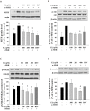
Ethyl linoleate is an unsaturated fatty acid used in many cosmetics for its various attributes, such as antibacterial and anti-inflammatory properties and clinically proven to be an effective anti-acne agent. In this study, we investigated the effect of ethyl linoleate on the melanogenesis and the mechanism underlying its action on melanogenesis in B16F10 murine melanoma cells. Our results revealed that ethyl linoleate significantly inhibited melanin content and intracellular tyrosinase activity in α-MSH-induced B16F10 cells, but it did not directly inhibit activity of mushroom tyrosinase. Ethyl linoleate inhibited the expression of microphthalmia-associated transcription factor (MITF), tyrosinase, and tyrosinase related protein 1 (TRP1) in governing melanin pigment synthesis. We observed that ethyl linoleate inhibited phosphorylation of Akt and glycogen synthase kinase 3β (GSK3β) and reduced the level of β-catenin, suggesting that ethyl linoleate inhibits melanogenesis through Akt/GSK3β/β-catenin signal pathway. Therefore, we propose that ethyl linoleate may be useful as a safe whitening agent in cosmetic and a potential therapeutic agent for reducing skin hyperpigmentation in clinics.
Keywords: Akt; Ethyl linoleate; GSK3β; Melanogenesis; β-catenin.
Conflict of interest statement
CONFLICTS OF INTEREST: The authors declare no conflicts of interest.
Figures


Similar articles
-
Sageretia thea fruit extracts rich in methyl linoleate and methyl linolenate downregulate melanogenesis via the Akt/GSK3β signaling pathway.Nutr Res Pract. 2018 Feb;12(1):3-12. doi: 10.4162/nrp.2018.12.1.3. Epub 2018 Jan 18. Nutr Res Pract. 2018. PMID: 29399291 Free PMC article.
-
Andrographolide suppresses melanin synthesis through Akt/GSK3β/β-catenin signal pathway.J Dermatol Sci. 2015 Jul;79(1):74-83. doi: 10.1016/j.jdermsci.2015.03.013. Epub 2015 Mar 30. J Dermatol Sci. 2015. PMID: 25869056
-
Sesamol Inhibited Melanogenesis by Regulating Melanin-Related Signal Transduction in B16F10 Cells.Int J Mol Sci. 2018 Apr 7;19(4):1108. doi: 10.3390/ijms19041108. Int J Mol Sci. 2018. PMID: 29642438 Free PMC article.
-
N-(4-bromophenethyl) Caffeamide Inhibits Melanogenesis by Regulating AKT/Glycogen Synthase Kinase 3 Beta/Microphthalmia-associated Transcription Factor and Tyrosinase-related Protein 1/Tyrosinase.Curr Pharm Biotechnol. 2015;16(12):1111-9. doi: 10.2174/1389201016666150817094258. Curr Pharm Biotechnol. 2015. PMID: 26278531
-
Neobavaisoflavone Inhibits Melanogenesis through the Regulation of Akt/GSK-3β and MEK/ERK Pathways in B16F10 Cells and a Reconstructed Human 3D Skin Model.Molecules. 2020 Jun 9;25(11):2683. doi: 10.3390/molecules25112683. Molecules. 2020. PMID: 32527040 Free PMC article.
Cited by
-
Purification of Ethyl Linoleate from Foxtail Millet (Setaria italica) Bran Oil via Urea Complexation and Molecular Distillation.Foods. 2021 Aug 19;10(8):1925. doi: 10.3390/foods10081925. Foods. 2021. PMID: 34441702 Free PMC article.
-
Phytochemical Characterization and Antioxidant Activity of Cajanus cajan Leaf Extracts for Nutraceutical Applications.Molecules. 2025 Apr 15;30(8):1773. doi: 10.3390/molecules30081773. Molecules. 2025. PMID: 40333766 Free PMC article.
-
Importance and Relevance of Phytochemicals Present in Galenia africana.Scientifica (Cairo). 2022 Jan 25;2022:5793436. doi: 10.1155/2022/5793436. eCollection 2022. Scientifica (Cairo). 2022. PMID: 35186343 Free PMC article. Review.
-
Evaluation of lipid oxidation mechanisms in beverages and cosmetics via analysis of lipid hydroperoxide isomers.Sci Rep. 2019 May 14;9(1):7387. doi: 10.1038/s41598-019-43645-1. Sci Rep. 2019. PMID: 31089240 Free PMC article.
-
Effects of Silybum marianum L. Seed Extracts on Multi Drug Resistant (MDR) Bacteria.Molecules. 2023 Dec 21;29(1):64. doi: 10.3390/molecules29010064. Molecules. 2023. PMID: 38202647 Free PMC article.
References
-
- Clarys P, Alewaeters K, Lambrecht R, Barel AO. Skin color measurements: comparison between three instruments: the Chromameter®, the DermaSpectrometer® and the Mexameter®. Skin Res Technol. 2000;6:230–238. - PubMed
-
- Sturm RA. Skin colour and skin cancer - MC1R, the genetic link. Melanoma Res. 2002;12:405–416. - PubMed
-
- Cui R, Widlund HR, Feige E, Lin JY, Wilensky DL, Igras VE, D'Orazio J, Fung CY, Schanbacher CF, Granter SR, Fisher DE. Central role of p53 in the suntan response and pathologic hyperpigmentation. Cell. 2007;128:853–864. - PubMed
LinkOut - more resources
Full Text Sources
Other Literature Sources
Research Materials

