Cilostazol attenuates kainic acid-induced hippocampal cell death
- PMID: 29302213
- PMCID: PMC5746513
- DOI: 10.4196/kjpp.2018.22.1.63
Cilostazol attenuates kainic acid-induced hippocampal cell death
Abstract
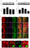
Cilostazol is a selective inhibitor of type 3 phosphodiesterase (PDE3) and has been widely used as an antiplatelet agent. Cilostazol mediates this activity through effects on the cyclic adenosine monophosphate (cAMP) signaling cascade. Recently, it has attracted attention as a neuroprotective agent. However, little is known about cilostazol's effect on excitotoxicity induced neuronal cell death. Therefore, this study evaluated the neuroprotective effect of cilostazol treatment against hippocampal neuronal damage in a mouse model of kainic acid (KA)-induced neuronal loss. Cilostazol pretreatment reduced KA-induced seizure scores and hippocampal neuron death. In addition, cilostazol pretreatment increased cAMP response element-binding protein (CREB) phosphorylation and decreased neuroinflammation. These observations suggest that cilostazol may have beneficial therapeutic effects on seizure activity and other neurological diseases associated with excitotoxicity.
Keywords: Cilostazol; Hippocampus; Kainic acid; Neuroinflammation; Neuronal death.
Conflict of interest statement
CONFLICTS OF INTEREST: The authors declare no conflicts of interest.
Figures


References
-
- Kimura Y, Tani T, Kanbe T, Watanabe K. Effect of cilostazol on platelet aggregation and experimental thrombosis. Arzneimittelforschung. 1985;35:1144–1149. - PubMed
-
- Kambayashi J, Liu Y, Sun B, Shakur Y, Yoshitake M, Czerwiec F. Cilostazol as a unique antithrombotic agent. Curr Pharm Des. 2003;9:2289–2302. - PubMed
-
- Miyamoto N, Tanaka R, Shimura H, Watanabe T, Mori H, Onodera M, Mochizuki H, Hattori N, Urabe T. Phosphodiesterase III inhibition promotes differentiation and survival of oligodendrocyte progenitors and enhances regeneration of ischemic white matter lesions in the adult mammalian brain. J Cereb Blood Flow Metab. 2010;30:299–310. - PMC - PubMed
-
- Lee JH, Park SY, Shin YW, Hong KW, Kim CD, Sung SM, Kim KY, Lee WS. Neuroprotection by cilostazol, a phosphodiesterase type 3 inhibitor, against apoptotic white matter changes in rat after chronic cerebral hypoperfusion. Brain Res. 2006;1082:182–191. - PubMed
-
- Qi DS, Tao JH, Zhang LQ, Li M, Wang M, Qu R, Zhang SC, Liu P, Liu F, Miu JC, Ma JY, Mei XY, Zhang F. Neuroprotection of Cilostazol against ischemia/reperfusion-induced cognitive deficits through inhibiting JNK3/caspase-3 by enhancing Akt1. Brain Res. 2016;1653:67–74. - PubMed
LinkOut - more resources
Full Text Sources
Other Literature Sources

