. 2017 May 23:2:14.
doi: 10.1038/s41536-017-0017-0. eCollection 2017.

Injection of embryonic stem cell derived macrophages ameliorates fibrosis in a murine model of liver injury

Affiliations

Injection of embryonic stem cell derived macrophages ameliorates fibrosis in a murine model of liver injury

Sharmin S Haideri et al. NPJ Regen Med. .

Erratum in

Abstract

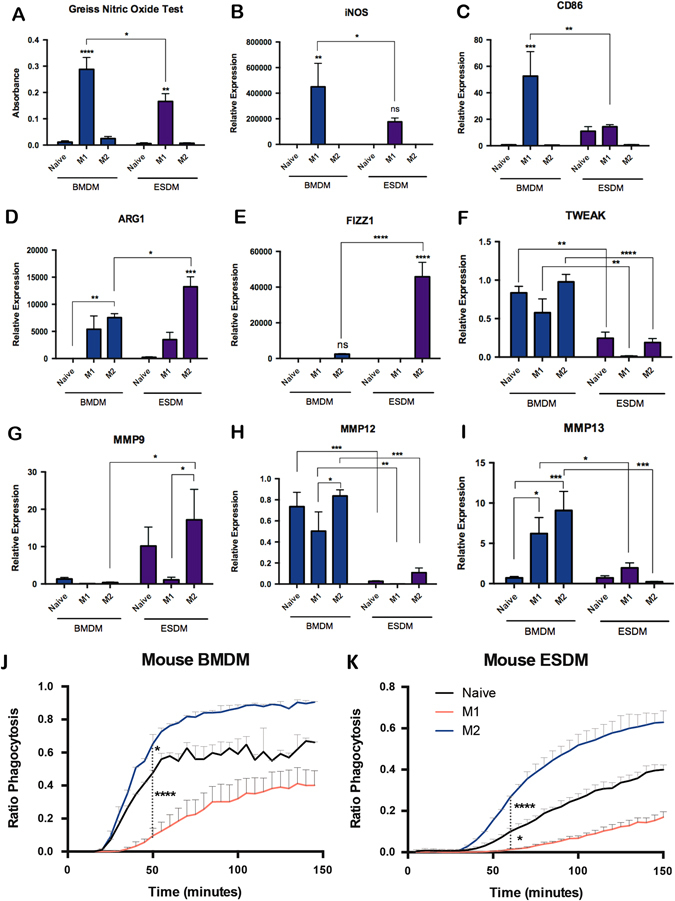
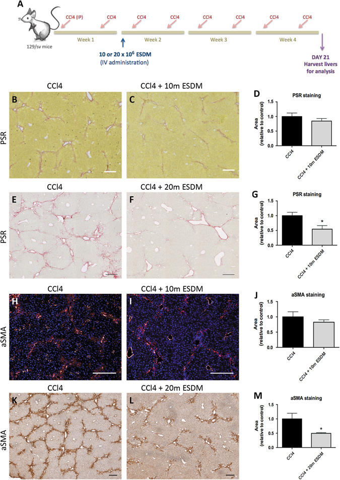
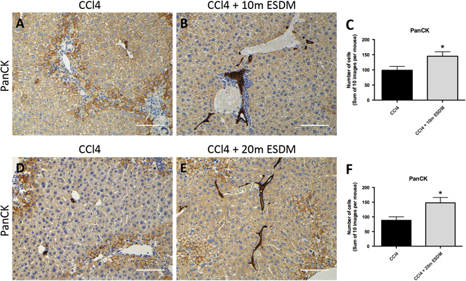
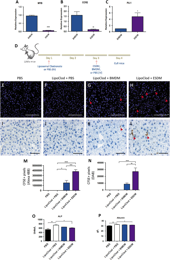
Chronic liver injury can be caused by viral hepatitis, alcohol, obesity, and metabolic disorders resulting in fibrosis, hepatic scarring, and cirrhosis. Novel therapies are urgently required and previous work has demonstrated that treatment with bone marrow derived macrophages can improve liver regeneration and reduce fibrosis in a murine model of hepatic injury and fibrosis. Here, we describe a protocol whereby pure populations of therapeutic macrophages can be produced in vitro from murine embryonic stem cells on a large scale. Embryonic stem cell derived macrophages display comparable morphology and cell surface markers to bone marrow derived macrophages but our novel imaging technique revealed that their phagocytic index was significantly lower. Differences were also observed in their response to classical induction protocols with embryonic stem cell derived macrophages having a reduced response to lipopolysaccharide and interferon gamma and an enhanced response to IL4 compared to bone marrow derived macrophages. When their therapeutic potential was assessed in a murine, carbon tetrachloride-induced injury and fibrosis model, embryonic stem cell derived macrophages significantly reduced the amount of hepatic fibrosis to 50% of controls, down-regulated the number of fibrogenic myofibroblasts and activated liver progenitor cells. To our knowledge, this is the first study that demonstrates a therapeutic effect of macrophages derived in vitro from pluripotent stem cells in a model of liver injury. We also found that embryonic stem cell derived macrophages repopulated the Kupffer cell compartment of clodronate-treated mice more efficiently than bone marrow derived macrophages, and expressed comparatively lower levels of Myb and Ccr2, indicating that their phenotype is more comparable to tissue-resident rather than monocyte-derived macrophages.


Conflict of interest statement

The authors declare that they have no competing financial interests.

Figures

Fig. 1
Fig. 1
Comparison of ESDMs and BMDMs. Stained cytospins of BMDMs (a) and ESDMs (b) with image analyses demonstrating that ESDMs are larger (c) (Scale bars 30 μM). Flow cytometry analyses of BMDMs (d) and ESDM (e) demonstrating a pure population in ESDMs. Images from live phagocytosis assay of BMDMs (g) and ESDMs (h) at 0 (I), 50 (II, 100 (III) and 150 (IV) minutes after the addition of Phrodo beads and quantification of rate of phagocytosis using Harmony image analysis software (i) (Scale bars 50 μM). See Supplementary Fig. S1 for details of the assay. [*p < 0.05, **p < 0.01, ***p < 0.001, ****p < 0.0001; n = 3, c Unpaired t-test, f One-way ANOVA with Kruskal-Wallis test, i Two-way ANOVA with Sidak’s multiple comparison test]
Fig. 2
Fig. 2
M1 and M2 stimulation of ESDMs and BMDMs. Griess nitric oxide assay of naïve, M1 and M2-stimulated BMDMs and ESDMs (a), and expression of key markers by qRT-PCR analyses, iNos (b) and Cd86 (c) as markers of M1, Arg1 (d) and Fizz1 (e) as markers of M2 phenotypes, and Tweak (f), Mmp9 (g), Mmp12 (h), and Mmp13 (i) as markers of tissue remodeling. Quantification of phagocytosis of M1-and M2-stimulated BMDMs (j) and ESDMs (k). [*p < 0.05, **p < 0.01, ***p < 0.001, ****p < 0.0001; n = 3 per group, Two-way ANOVA with Tukey’s multiple comparison test]
Fig. 3
Fig. 3
Production of macrophages from human PSCs. Macrophage derived from hPSC express both CD11b and 25F9 (b) but not CD93 (a) and key M1 (c, d) and M2-associated genes (e, f) are upregulated upon stimulation. Assessed using a live imaging phagocytosis assay, naïve and M2-activated macrophages were more phagocytic than M1-activated macrophages and this was observed for both MDMs (g) and iPSC-DMs (h). [*p < 0.05, **p < 0.01, ***p < 0.001; n = 3, cf One-way ANOVA, g, h Two-way ANOVA with Tukey’s multiple comparison test]
Fig. 4
Fig. 4
The higher dose of ESDMs has an anti-fibrotic effect. Schematic a of injury and macrophage injection of 10 or 20 million (m) macrophages. PSR staining (bg) and aSMA staining (hm) of liver sections from CCl4-treated livers that were injected with either 10 million (10 m) or 20 million (20 m) ESDMs. Scale bars 200 μM. [*p < 0.05, n = 5 per group, Unpaired t-test]
Fig. 5
Fig. 5
ESDMs can initiate a progenitor response in injured livers. A significantly higher number of PanCK-positive cells were observed in mice injected with ESDMs compared to control injury livers (af). Scale bars 100 μM. [*p < 0.05, n = 5 per group, Unpaired t-test]
Fig. 6
Fig. 6
ESDMs can repopulation the liver of liposomal chlodronate-treated mice. Expression of markers differentially expressed in monocyte-derived vs. tissue-resident macrophages, Myb (a), Ccr2 (b), and Pu.1 (c). Schematic representation (d) of liposomal clodronate-induced Kupffer cell depletion and injection of ESDM or BMDM. CFSE immunostaining (eh Scale bars 100 μM) and CFSE immunohistochemistry (il Scale bars 50 μM.) of liver sections from the different treatment groups. Quantification of CFSE+ immunostaining (m) and CFSE+ immunohistochemistry (n). Arrowheads point at CFSE+ cells detected. Blood serum analysis of o Alkaline phosphatase (ALP), and p Albumin levels. [*p < 0.05, **p < 0.01, ***p < 0.001, ****p < 0.0001; n = 5 per group, One-way ANOVA]

Comment in

References

    1. Forbes SJ, Rosenthal N. Preparing the ground for tissue regeneration: from mechanism to therapy. Nat. Med. 2014;20:857–869. doi: 10.1038/nm.3653. - DOI - PubMed
    1. Friedman SL. Mechanisms of hepatic fibrogenesis. Gastroenterology. 2008;134:1655–1669. doi: 10.1053/j.gastro.2008.03.003. - DOI - PMC - PubMed
    1. Soltys KA, et al. Barriers to the successful treatment of liver disease by hepatocyte transplantation. J. Hepatol. 2010;53:769–774. doi: 10.1016/j.jhep.2010.05.010. - DOI - PMC - PubMed
    1. Sakaida I, et al. Transplantation of bone marrow cells reduces CCl4-induced liver fibrosis in mice. Hepatology. 2004;40:1304–1311. doi: 10.1002/hep.20452. - DOI - PubMed
    1. Thomas JA, et al. Macrophage therapy for murine liver fibrosis recruits host effector cells improving fibrosis, regeneration, and function. Hepatology. 2011;53:2003–2015. doi: 10.1002/hep.24315. - DOI - PubMed