Review
. 2017 Jun 16:2:18.
doi: 10.1038/s41536-017-0023-2. eCollection 2017.

Biomaterial-driven in situ cardiovascular tissue engineering-a multi-disciplinary perspective

Affiliations
Review

Biomaterial-driven in situ cardiovascular tissue engineering-a multi-disciplinary perspective

Tamar B Wissing et al. NPJ Regen Med. .

Abstract

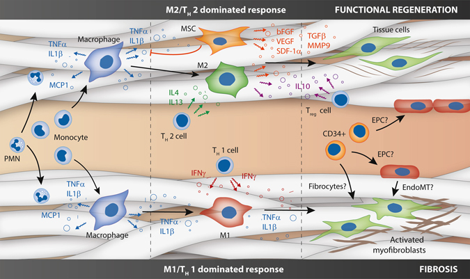
There is a persistent and growing clinical need for readily-available substitutes for heart valves and small-diameter blood vessels. In situ tissue engineering is emerging as a disruptive new technology, providing ready-to-use biodegradable, cell-free constructs which are designed to induce regeneration upon implantation, directly in the functional site. The induced regenerative process hinges around the host response to the implanted biomaterial and the interplay between immune cells, stem/progenitor cell and tissue cells in the microenvironment provided by the scaffold in the hemodynamic environment. Recapitulating the complex tissue microstructure and function of cardiovascular tissues is a highly challenging target. Therein the scaffold plays an instructive role, providing the microenvironment that attracts and harbors host cells, modulating the inflammatory response, and acting as a temporal roadmap for new tissue to be formed. Moreover, the biomechanical loads imposed by the hemodynamic environment play a pivotal role. Here, we provide a multidisciplinary view on in situ cardiovascular tissue engineering using synthetic scaffolds; starting from the state-of-the art, the principles of the biomaterial-driven host response and wound healing and the cellular players involved, toward the impact of the biomechanical, physical, and biochemical microenvironmental cues that are given by the scaffold design. To conclude, we pinpoint and further address the main current challenges for in situ cardiovascular regeneration, namely the achievement of tissue homeostasis, the development of predictive models for long-term performances of the implanted grafts, and the necessity for stratification for successful clinical translation.


Conflict of interest statement

Prof. Bouten is shareholder of Xeltis BV. Ms. Wissing, Ms. Bonito and Dr. Smits declare no potential competing financial interests.

Figures

Fig. 1
Fig. 1
Overview of the different stages of in situ tissue regeneration, going from a synthetic, biodegradable bare construct toward a viable substitute (a). Although many aspects underlying in situ regeneration remain unknown, it is hypothesized that the stages mirror the natural phases of the wound healing response (b), starting with the inflammatory phase, characterized by the infiltration of immune cells and the formation of a preliminary matrix. The subsequent proliferative phase is characterized by a secondary influx of immune and tissue producing cells, extracellular matrix (ECM) deposition, angiogenesis and (re-)endothelialization of the construct. Tissue homeostasis is restored after a remodeling phase of the newly formed ECM and the resolution of inflammation. Photographs of heart valves adapted from; photographs of vascular grafts courtesy of Renée Duijvelshoff
Fig. 2
Fig. 2
Cartoon of the various (simplified) cell-cell interactions in in situ tissue engineering, as hypothesized based on the state-of-the-art. After the instantaneous response of protein adhesion and platelet activation (not depicted), circulating polymorphonuclear cells and monocytes are recruited to an implanted scaffold in response to various chemokines (e.g., monocyte chemoattractant protein (MCP)-1). Upon activation, the monocytes give rise to macrophages in the scaffold, which are a source of pro-inflammatory factors (e.g., tumor necrosis factor (TNF)-α, Interleukin (IL)‐1β). Depending on the scaffold properties, this is followed by an M1/TH 1 cell dominated response pro-inflammatory response (bottom) of an M2/TH 2 cell dominated pro-regenerative response (top). The former is characterized by the prolonged presence of M1 macrophages, instigated by TH1 cell-secreted pro-inflammatory cytokines, such as interferon-γ. Recruited fibroblasts typically acquire an activated phenotype, producing non-functional cross-linked fibrous scar tissue. In contrast, the pro-regenerative process is dominated by M2 macrophages under influence of TH2 cell secreted cytokines (e.g., interleukin (IL)-4 and -13). Mesenchymal stromal cells play an important immunomodulatory role by inhibiting pro-inflammatory factors, such as TNF-α, as well as secreting numerous trophic factors (e.g., basic fibroblast growth factor, bFGF; vascular endothelial growth factor, VEGF; stromal cell-derived factor-1α, SDF-1α; transforming growth factor β, TGF-β; matrix metalloproteinase 9, MMP-9). This biochemical milieu attracts tissue cells and modulates the formation and remodeling of well-organized functional neotissue. Upon scaffold degradation, T reg cells inhibit the inflammatory process by secretion of, e.g., IL-10. Homing of circulatory CD34+ progenitor cells, such as fibrocytes and endothelial progenitor cells, as well as endothelial-to-mesenchymal transformation may contribute to cellularization and pathophysiological neotissue formation, although these processes are topic of active debate
Fig. 3
Fig. 3
Schematic representation of the design strategies that can be employed to tailor resident cell behavior inside the graft. The transfer of hemodynamic loads (a) can be tuned via adaptations in material properties, such as the mechanical properties, geometry (b) and microstructure (d). Concurrently, cell behavior is defined by interdependent microstructural parameters (e.g., fiber diameter, alignment, pore size and topography; (d) and biochemical parameters (e.g., surface chemistry and bio-activation); (c). However, local loads and scaffold parameters change in time due to material degradation (e) and new tissue formation. Subfigures b and e are adapted from refs. and , respectively
Fig. 4
Fig. 4
a Schematic illustration displaying the three main interdependent challenges faced for successful, robust in-man application of in situ tissue engineered cardiovascular grafts. The development process is represented by a continuous feedback loop between the optimization of the graft design and the development of predictive models to understand and determine long-term in vivo performance, while taking into account graft recipient variability (e.g., age, gender, co-morbidities, and utility). b Optimization of graft design is visualized as a flowchart, in which interchangeable scaffold design parameters together with the hemodynamic loads and cells will determine if tissue homeostasis will be reached. Societal demands, including patient and physician wishes, should be taken into consideration during the (early) stages of graft development to determine the added value of these grafts for health care. HTA: Health Technology Assessment

References

    1. Mol A, Smits AIPM, Bouten CVC, Baaijens FPT. Tissue engineering of heart valves: advances and current challenges. Expert. Rev. Med. Devices. 2009;6:259–275. doi: 10.1586/erd.09.12. - DOI - PubMed
    1. Bouten CVC, et al. Substrates for cardiovascular tissue engineering. Adv. Drug. Deliv. Rev. 2011;63:221–241. doi: 10.1016/j.addr.2011.01.007. - DOI - PubMed
    1. van Loon, S. L. M., Smits, A. I. P. M., Driessen-Mol, A., Baaijens, F. P. T. & Bouten, C. V. C. The Immune Response in In Situ Tissue Engineering of Aortic Heart Valves. Calcific Aortic Valve Disease (ed. Aikawa, E.) 207–245 (2013).
    1. MacGrogan, D. et al. How to make a heart valve: from embryonic development to bioengineering of living valve substitutes. Cold Spring Harb. Perspect. Med. 4, a013912 (2014). - PMC - PubMed
    1. Schoen FJ. Evolving concepts of cardiac valve dynamics: the continuum of development, functional structure, pathobiology, and tissue engineering. Circulation. 2008;118:1864–1880. doi: 10.1161/CIRCULATIONAHA.108.805911. - DOI - PubMed