. 2017 Dec 15:2:32.
doi: 10.1038/s41536-017-0037-9. eCollection 2017.

Intraoperative delivery of the Notch ligand Jagged-1 regenerates appendicular and craniofacial bone defects

Affiliations

Intraoperative delivery of the Notch ligand Jagged-1 regenerates appendicular and craniofacial bone defects

Daniel W Youngstrom et al. NPJ Regen Med. .

Abstract

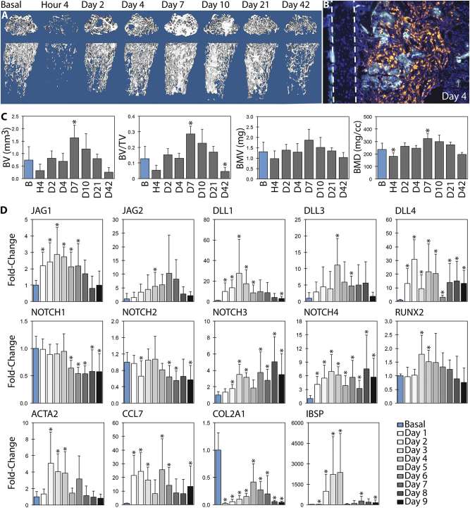
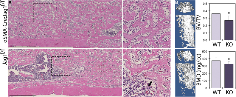
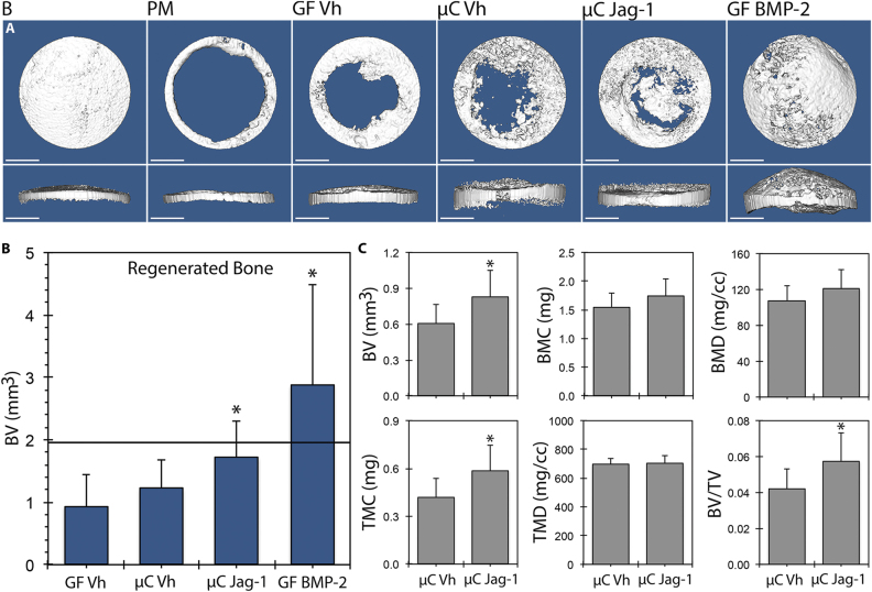
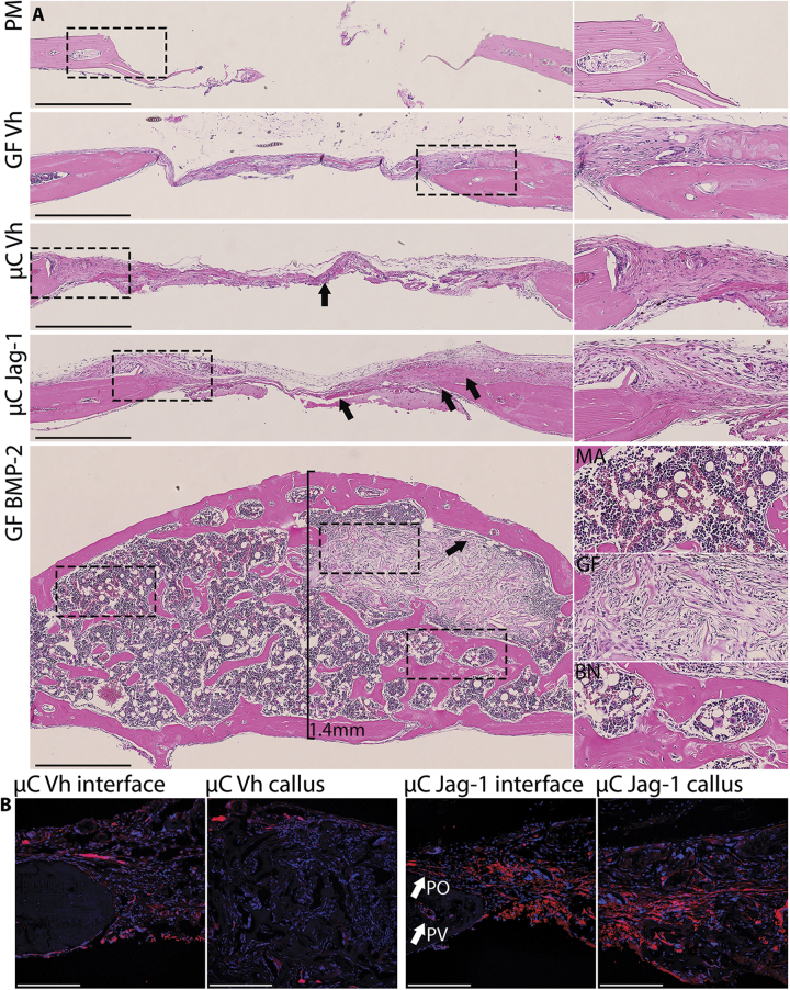
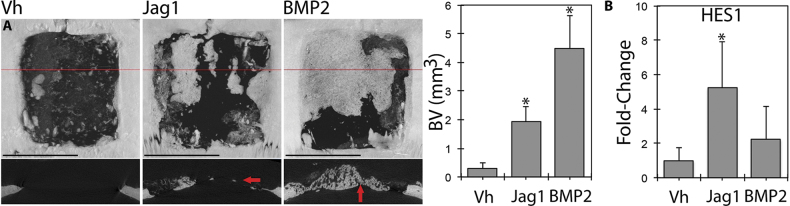
Each year, 33% of US citizens suffer from a musculoskeletal condition that requires medical intervention, with direct medical costs approaching $1 trillion USD per year. Despite the ubiquity of skeletal dysfunction, there are currently limited safe and efficacious bone growth factors in clinical use. Notch is a cell-cell communication pathway that regulates self-renewal and differentiation within the mesenchymal/osteoblast lineage. The principal Notch ligand in bone, Jagged-1, is a potent osteoinductive protein that positively regulates post-traumatic bone healing in animals. This report describes the temporal regulation of Notch during intramembranous bone formation using marrow ablation as a model system and demonstrates decreased bone formation following disruption of Jagged-1 in mesenchymal progenitor cells. Notch gain-of-function using recombinant Jagged-1 protein on collagen scaffolds promotes healing of craniofacial (calvarial) and appendicular (femoral) surgical defects in both mice and rats. Localized delivery of Jagged-1 promotes bone apposition and defect healing, while avoiding the diffuse bone hypertrophy characteristic of the clinically problematic bone morphogenetic proteins. It is concluded that Jagged-1 is a bone-anabolic agent with therapeutic potential for regenerating traumatic or congenital bone defects.


Conflict of interest statement

K.D.H. is the co-founder of Skelegen, LLC and holds US patent application 14/760,371. Other authors declare no competing financial interests.

Figures

Fig. 1
Fig. 1
µCT and qPCR profile post-marrow ablation. Mechanical disruption of the marrow environment results in deposition of intramedullary bone within seven days. This was quantified by µCT (n = 6), with representative sagittal and coronal 3D reconstructions shown (a, c). Osteoblasts derived from αSMA-positive mesenchymal progenitor cells produce new (double-labeled) bone: the cortical wall is demarcated with dashed lines (b). Expression of Notch ligands, receptors, and select marker genes are presented over time by qPCR with respect to basal bone, relative to ACTB (d). Notch ligands are upregulated during osteoblast differentiation. Statistical significance is symbolized with * for p < 0.05 (n = 6–12)
Fig. 2
Fig. 2
Jag1-KO reduces post-ablation bone formation. Cre-positive and Cre-negative female littermates in the Jag1f/f line (n = 3) underwent marrow ablation procedures and received single tamoxifen injections at time of surgery. H&E histology (a) is shown alongside µCT reconstructions and quantitation (b) for day 7 post ablation. Jag1-KO animals underwent incomplete osteoblastogenesis and contain regions of marrow interspersed within the newly formed trabecular bone (arrow). Scale bars are equal to 500 µm. Statistical significance is symbolized with * for p < 0.05
Fig. 3
Fig. 3
Jag1 improves healing of calvarial defects in mice. 3D reconstructions (a) and total regenerated bone (b) (PM-subtracted) were assessed using µCT 42 days post-defect, reported at a threshold of 666. Total ROI quantitation between µC-Vh and µC-Jag1 groups (c) was conducted at a threshold of 1650. Statistical significance is symbolized with * for p < 0.05 (n = 8–12)
Fig. 4
Fig. 4
Jag1 activates αSMA-positive mesenchyme in the calvaria without bone overgrowth. Representative H&E histology (a) shows increased periosteal mesenchyme and improved bone matrix production 42 days post defect in the µC-Jag1 group (arrows) relative to the incomplete mineralization and fibrosis in the associated µC-Vh control (arrow). BMP2 induced supraphysiological production of bone (BN) and fatty marrow (MA), encapsulating and preserving the Gelfoam scaffold (GF). Select ROIs are zoomed adjacent to source images and marked using dashed lines. Scale bars are equal to 500 µm. αSMA-CreERT2;tdTomato reporter activity is increased in the µC-Jag1 group relative to its µC-Vh control (b). Confocal scale bars are equal to 200 µm
Fig. 5
Fig. 5
Jag1 promotes healing of femoral defects. Representative H&E histology (a) shows improved healing of Jag1-treated defects relative to contralateral GF-Vh controls. Selected ROIs are zoomed adjacent to source images and marked using dashed lines. At day 10, local regions of endochondral ossification occur adjacent to residual Gelfoam and the cortical margin (arrow). The Jag1-induced increase in intramedullary and cortical bone is evident at day 20 (arrows). Scale bars are equal to 500 µm. µCT isosurfaces and data b were gathered at a threshold of 2667. Statistical significance is symbolized with * for p < 0.05 (n = 5). Representative images represent matched sets from individual animals
Fig. 6
Fig. 6
Jag1 regenerates critical-sized calvarial defects in rats. Jag1 drives pronounced bone healing in rat segmental skull defects, as seen via µCT (a). Representative dorsal 3D reconstructions are shown with 2D transverse sections (red lines) below. Jag1-induced healing occurs within the plane of surrounding bone (arrow), while BMP2 causes bone overgrowth above the injury margin: termed hypertrophy (arrow). Expression of the canonical Notch target gene, HES1, is increased in a frontal quadrant of rat calveriae relative to GAPDH in the Jag1-treated animals preceding this bone formation in the rostral portion of the healing defect (b). Associated quantitation represents bone within 6.6 × 6.6 mm ROIs centered in each defect. Scale bars are equal to 5 mm. Statistical significance is symbolized with * for p < 0.05 (n = 4–6)

References

    1. van Staa TP, Dennison EM, Leufkens HG, Cooper C. Epidemiology of fractures in England and Wales. Bone. 2001;29:517–522. doi: 10.1016/S8756-3282(01)00614-7. - DOI - PubMed
    1. Antonova E, Le TK, Burge R, Mershon J. Tibia shaft fractures: costly burden of nonunions. BMC Musculoskelet. Disord. 2013;14:42. doi: 10.1186/1471-2474-14-42. - DOI - PMC - PubMed
    1. Mankin HJ, Gebhardt MC, Jennings LC, Springfield DS, Tomford WW. Long-term results of allograft replacement in the management of bone tumors. Clin. Orthop. Relat. Res. 1996;324:86–97. doi: 10.1097/00003086-199603000-00011. - DOI - PubMed
    1. Burge R, et al. Incidence and economic burden of osteoporosis-related fractures in the United States, 2005-2025. J. Bone Miner. Res. 2007;22:465–475. doi: 10.1359/jbmr.061113. - DOI - PubMed
    1. Nikkel LE, et al. Impact of comorbidities on hospitalization costs following hip fracture. J. Bone Jt. Surg. 2012;94:9–17. doi: 10.2106/JBJS.J.01077. - DOI - PubMed

LinkOut - more resources