. 2017 Dec;6(6):611-619.
doi: 10.21037/gs.2017.10.03.

Robotic and endoscopic transoral thyroidectomy: feasibility and description of the technique in the cadaveric model

Affiliations

Robotic and endoscopic transoral thyroidectomy: feasibility and description of the technique in the cadaveric model

Bora Kahramangil et al. Gland Surg. 2017 Dec.

Abstract

Background: Numerous new approaches have been described over the years to improve the cosmetic outcomes of thyroid surgery. Transoral approach is a new technique that aims to achieve superior cosmetic outcomes by concealing the incision in the oral cavity.

Methods: Transoral thyroidectomy through vestibular approach was performed in two institutions on cadaveric models. Procedure was performed endoscopically in one institution, while the robotic technique was utilized at the other.

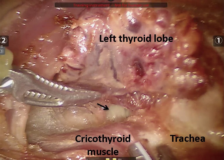
Results: Transoral thyroidectomy was successfully performed at both institutions with robotic and endoscopic techniques. All vital structures were identified and preserved.

Conclusions: Transoral thyroidectomy has been performed in animal and cadaveric models, as well as in some clinical studies. Our initial experience indicates the feasibility of this approach. More clinical studies are required to elucidate its full utility.

Keywords: Robotic; endoscopic; thyroidectomy; transoral.


Conflict of interest statement

Conflicts of Interest: The authors have no conflicts of interest to declare.

Figures

Figure 1
Figure 1
Intraoperative photos describing the placement of incisions and trocars in the robotic technique. (A) A midline incision was made for the robotic camera (arrow), and two lateral incisions for the robotic instruments (arrowheads); (B) the camera port was inserted through the midline incision and the ports for robotic instruments through the lateral incisions.
Figure 2
Figure 2
Intraoperative photos demonstrating docking and robotic instruments used for transoral thyroidectomy. (A) Robot is brought in and docked from the head of the bed; (B) a 30-degree down-looking camera is introduced from the middle port. A robotic Maryland forceps is inserted from the left port and a Harmonic scalpel from the right.
Figure 3
Figure 3
Intraoperative capture showing the use of traction sutures during robotic procedure. Simultaneous use of traction sutures and insufflation resulted in better exposure compared to insufflation alone.
Figure 4
Figure 4
Intraoperative photo showing the robotic resection of the left thyroid lobe. During resection, the left recurrent laryngeal nerve (arrow) was identified and preserved.
Figure 5
Figure 5
Intraoperative photo demonstrating the right thyroid lobe dissection. The right lobe is retracted to the left while the tissue attachments are dissected using the Harmonic scalpel (on the right side).
Figure 6
Figure 6
Intraoperative photo after completion of robotic dissection of the thyroid. The thyroid was divided at the isthmus prior to dissection. The right and left lobes can be seen on both sides of the picture. Specimens were laparoscopically removed through the midline incision.
Figure 7
Figure 7
Intraoperative photos describing the placement of incisions and trocars in the endoscopic technique. (A) A 10-mm midline incision was used for the endoscopic camera, and two lateral 5-mm incisions for the endoscopic instruments. The courses of the bilateral lingual nerves are depicted in white; (B) the midline trocar (blue) was used for the endoscopic camera and specimen extraction. Endoscopic instruments were introduced through the lateral ports (red).
Figure 8
Figure 8
Postoperative photo showing the successfully removed thyroid gland using the endoscopic technique.
Figure 9
Figure 9
Intraoperative capture demonstrating the bilateral mental nerves (red circles) that were identified and preserved during endoscopic resection.
Figure 10
Figure 10
Immediate postoperative capture after endoscopic resection showing the cervical skin. The mentum and the anterior cervical skin were noted to retain their original elasticity and appearance.

References

    1. Jeannon JP, Orabi AA, Bruch GA, et al. Diagnosis of recurrent laryngeal nerve palsy after thyroidectomy: a systematic review. Int J Clin Pract 2009;63:624-9. 10.1111/j.1742-1241.2008.01875.x - DOI - PubMed
    1. Almquist M, Hallgrimsson P, Nordenstrom E, et al. Prediction of permanent hypoparathyroidism after total thyroidectomy. World J Surg 2014;38:2613-20. 10.1007/s00268-014-2622-z - DOI - PubMed
    1. Arora A, Swords C, Garas G, et al. The perception of scar cosmesis following thyroid and parathyroid surgery: a prospective cohort study. Int J Surg 2016;25:38-43. 10.1016/j.ijsu.2015.11.021 - DOI - PubMed
    1. Lee S, Kim HY, Lee CR, et al. A prospective comparison of patient body image after robotic thyroidectomy and conventional open thyroidectomy in patients with papillary thyroid carcinoma. Surgery 2014;156:117-25. 10.1016/j.surg.2014.02.007 - DOI - PubMed
    1. Miccoli P, Berti P, Conte M, et al. Minimally invasive surgery for thyroid small nodules: preliminary report. J Endocrinol Invest 1999;22:849-51. 10.1007/BF03343657 - DOI - PubMed

LinkOut - more resources