Review
. 2018 Jun;91(1086):20170725.
doi: 10.1259/bjr.20170725. Epub 2018 Jan 17.

The management of deep-seated, lowgrade lipomatous lesions

Affiliations
Review

The management of deep-seated, lowgrade lipomatous lesions

Zeid Al-Ani et al. Br J Radiol. 2018 Jun.

Abstract

Deep-seated, low-grade lipomatous lesions detected on imaging often cause uncertainty for diagnosis and treatment. Confidently distinguishing lipomas from well-differentiated liposarcomas is often not possible on imaging. The approach to management of such lesions varies widely between institutions. Applying an evidenced-based approach set around published literature that clearly highlights how criteria such as lesion size, location, age and imaging features can be used to predict the risk of well-differentiated liposarcomas and subsequent de-differentiation would seem sensible. Our aim is to review the literature and produce a unified, evidence-based guideline that will be a useful tool for managing these lesions.


Figures

Figure 1.
Figure 1.
Intramuscular lipoma within the flexor compartment of the forearm. (a) Axial T1 weighted image shows only thin septations but no thick septa or nodules. (b) Axial T2 fat suppressed image shows homogenous fat suppression without appreciable septa or non-fatty component.
Figure 2.
Figure 2.
Intramuscular liposarcoma within the thigh (vastus lateralis muscle). (a) Axial T1 weighted image and (b) axial T2 fat suppressed image showing a simple, predominantly fatty intramuscular lipomatous lesion with a few intervening muscle fibres but no thick septa or nodules. Percutaneous biopsy was performed with immunohistochemistry assessment confirming this as a WDL.
Figure 3.
Figure 3.
Intramuscular liposarcoma in the right thigh. (a) Sagittal T1 weighted image demonstrates thick septa (arrow) and nodular component (arrow head). (b) Sagittal T2 fat suppressed image demonstrates the non-fatty component as high signal septa (arrow) and nodule (arrow head).
Figure 4.
Figure 4.
Spindle cell lipoma variant in the paraspinal muscles. (a) Sagittal T1 weighted image and (b) sagittal T2 fat suppressed image demonstrate mixture of fatty (arrows) and non-fatty components (arrow heads) with incomplete signal suppression on the fat saturated image.
Figure 5.
Figure 5.
Intramuscular lipoma. Coronal T1 weighted image shows the characteristic inter-digitating margins (arrows) of an intramuscular lipoma within the obturator externus muscle.
Figure 6.
Figure 6.
Retroperitoneal liposarcoma. Axial contrast enhanced CT abdomen and pelvis examination demonstrates a large retroperitoneal fatty mass (arrows) with more complex, solid areas internally. The mass is significantly displacing bowel loops.
Figure 7.
Figure 7.
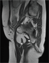
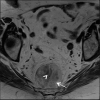
Presacral myelolipoma. Axial T1 weighted image demonstrates the fatty (arrow) and non-fatty haematopoietic (arrow head) components within the lesion in a typical presacral position.
Figure 8.
Figure 8.
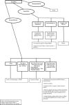
Suggested pathway for management of deep lipomatous lesions.

References

    1. Rydholm A, Berg NO, Size BNO. Size, site and clinical incidence of lipoma. Factors in the differential diagnosis of lipoma and sarcoma. Acta Orthop Scand 1983; 54: 929–34. doi: 10.3109/17453678308992936 - DOI - PubMed
    1. Fletcher CDM, Bridge JA, Hogendoorn P, Mertens F. WHO classification of tumours. In: International Agency for Research on Cancer (IARC) WHO classification of tumours. Volume ; 2013. pp. 19–46.
    1. Kempson RR, Fletcher CD, Evans HL, Hendrickson RM, Sibley R. Malignant lipomatous tumors. In: Atlas of tumor pathology: tumor of the soft tissue: The British Institute of Radiology.; 2001. pp. 217–38.
    1. Dalal KM, Antonescu CR, Singer S. Diagnosis and management of lipomatous tumors. J Surg Oncol 2008; 97: 298–313. doi: 10.1002/jso.20975 - DOI - PubMed
    1. Murphey MD, Arcara LK, Fanburg-Smith J. From the archives of the AFIP: imaging of musculoskeletal liposarcoma with radiologic-pathologic correlation. Radiographics 2005; 25: 1371. doi: 10.1148/rg.255055106 - DOI - PubMed

MeSH terms

LinkOut - more resources