. 2018 Jan 5;13(1):e0181529.
doi: 10.1371/journal.pone.0181529. eCollection 2018.

Tumor targeted delivery of doxorubicin in malignant peripheral nerve sheath tumors

Affiliations

Tumor targeted delivery of doxorubicin in malignant peripheral nerve sheath tumors

A B Madhankumar et al. PLoS One. .

Abstract

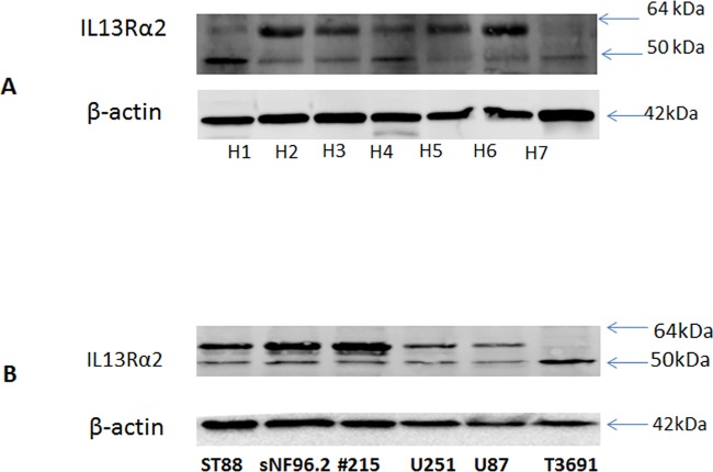
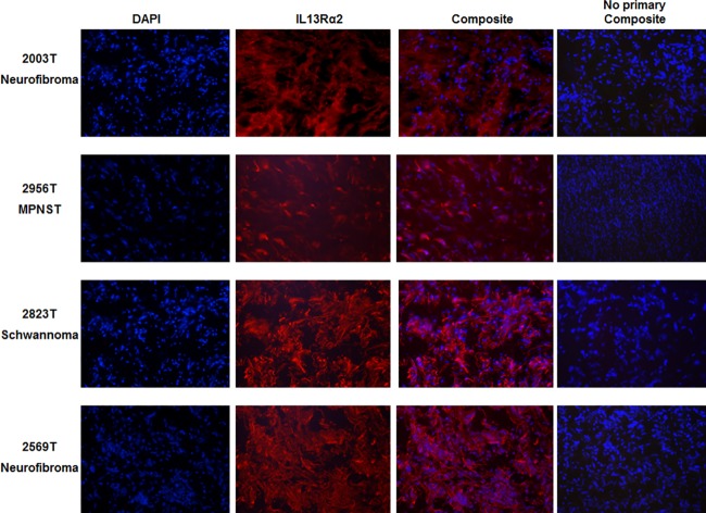
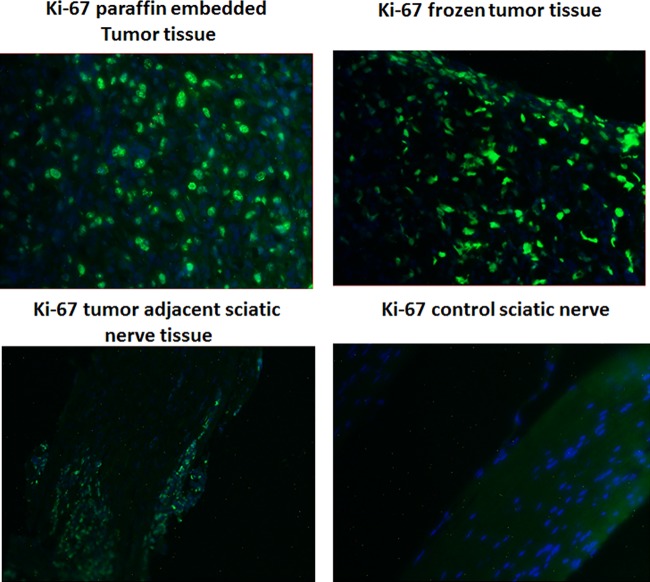
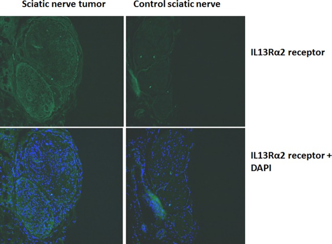
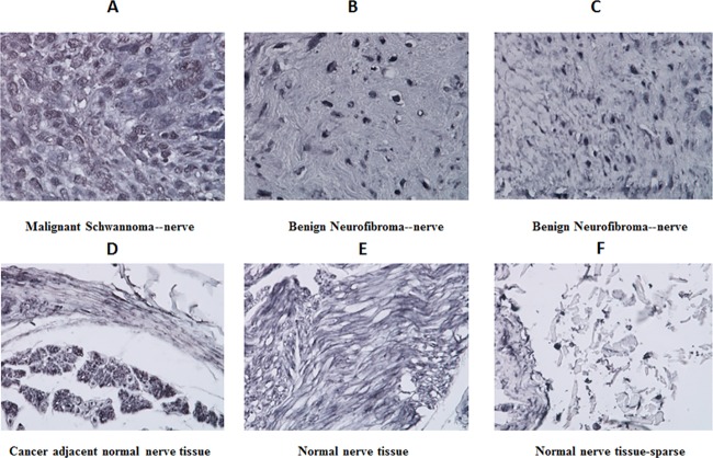
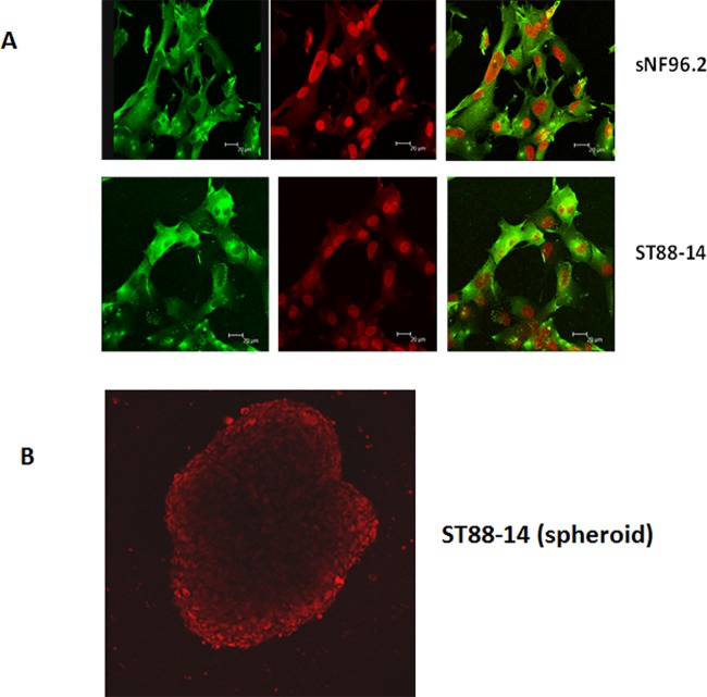
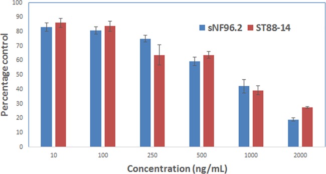
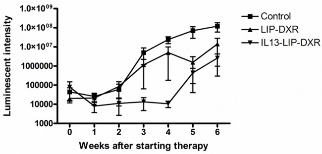
Peripheral nerve sheath tumors are benign tumors that have the potential to transform into malignant peripheral nerve sheath tumors (MPNSTs). Interleukin-13 receptor alpha 2 (IL13Rα2) is a cancer associated receptor expressed in glioblastoma and other invasive cancers. We analyzed IL13Rα2 expression in several MPNST cell lines including the STS26T cell line, as well as in several peripheral nerve sheath tumors to utilize the IL13Rα2 receptor as a target for therapy. In our studies, we demonstrated the selective expression of IL13Rα2 in several peripheral nerve sheath tumors by immunohistochemistry (IHC) and immunoblots. We established a sciatic nerve MPNST mouse model in NIH III nude mice using a luciferase transfected STS26T MPNST cell line. Similarly, analysis of the mouse sciatic nerves after tumor induction revealed significant expression of IL13Rα2 by IHC when compared to a normal sciatic nerve. IL13 conjugated liposomal doxorubicin was formulated and shown to bind and internalized in the MPNST cell culture model demonstrating cytotoxic effect. Our subsequent in vivo investigation in the STS26T MPNST sciatic nerve tumor model indicated that IL13 conjugated liposomal doxorubicin (IL13LIPDXR) was more effective in inhibiting tumor progression compared to unconjugated liposomal doxorubicin (LIPDXR). This further supports that IL13 receptor targeted nanoliposomes is a potential approach for treating MPNSTs.


Conflict of interest statement

Competing Interests: The authors have declared that no competing interests exist.

Figures

Fig 1
Fig 1
A. Immunoblots showing the expression levels of IL13Rα2 in human tissue homogenates from various nerve tumor tissues: H1- schwannoma, H2-neurofibroma, H3-schwannoma, H4-MPNST, H5-schwannoma, H6-neurofibroma, H7-neurofibroma. B. Human neurofibroma cell lines sNF96.2, ST88-14 and #215 cell lines were all found to express IL13Rα2 as evidenced from the Western blots.
Fig 2
Fig 2. Immunofluorescence of benign and malignant peripheral nerve tumors for IL13Rα2 (red).
The blue color represents the nuclear staining with DAPI. IL13Rα2 expression is evident in MPNST, low grade schwannomas and neurofibromas. From the composite images, most of the staining appears to be cytoplasmic as well as nuclear.
Fig 3
Fig 3. Immunohistochemistry for Ki-67 expression on the sciatic nerves from the tumor induced and control (untreated) mice indicates that the IL13Rα2 staining pattern was intense in tumor tissue.
Fig 4
Fig 4. IL13Rα2 expression in the sciatic nerve tumor developed in NIHIII mice injected with sNF96.2 NF-1 cells.
The expression levels were compared with the control sciatic nerves which were not injected with the cells.
Fig 5
Fig 5. IL13Rα2 expression in various tissues from a peripheral nerve carcinoma tissue microarray.
Representative microscopic images from the tissue microarray after IHC for IL13Rα2 expression are shown (panel A-F). Malignant Schwannoma distinctly shows robust expression of IL13Rα2 in the tissue (D) with positive expression in benign neurofibromas as evident from the IHC (E,F). Normal nerve tissue indicated minimal staining without distinct cellular morphology. No IL13Rα2 staining was observed in cancer adjacent normal tissues.
Fig 6
Fig 6. Fluorescent microscopic images of sNF96.2 and ST88-14 MPNST cells after exposure to IL13LIPDXR for 2 hours, followed by immunocytochemistry for IL13Rα2.
The green color in the cytoplasmic region represents the expression of IL13Rα2 receptor and the red color represents the endogenous fluorescence of the internalized doxorubicin delivered through targeted liposomes. (B) IL13LIPDXR is also able to bind and internalize through multicellular spheroids after exposure for 2 hours as evidenced by confocal microscopy images. More details can be found in the S2 Fig and S1 Video.
Fig 7
Fig 7. Cell proliferation assay performed on sNF96.2 and ST88-14 MPNST cells cultured in monolayer after treatment with IL13LIPDXR.
The results are displayed as percentage control with respect to untreated cells. 48 hours post treatment the cells were stained with Alamar blue (resazurin), a redox dye which exploits the reducing potential of the viable cells, whereby the non-fluorescent resazurin will be converted to a highly fluorescent resorufin, which can be monitored by a fluorescent plate reader.
Fig 8
Fig 8
In vivo tumor progression in the murine MPNST tumor model after administration of a once per week dose of 7 mg/kg body weight of (a) IL13LIPDXR (b) unconjugated LIPDXR and (c) control mice which were injected with saline.
Fig 9
Fig 9. Serum chemistry analysis.
The analysis was performed 48 hours post injection of the 7mg/kg dose of targeted and non-targeted liposomal doxorubicin. The values were compared with control mice injected with phosphate buffered saline. The levels indicate that liver functional enzymes including bilirubin and alkaline phosphatase are comparable in both the treated and control groups of mice. Creatinine and BUN levels are also comparable to that of untreated control mice, indicating that renal function is not significantly affected due to treatment.

Similar articles

Cited by

References

    1. Ferrari A, Bisogno G, Macaluso A, Casanova M, D'Angelo P, Pierani P, et al. Soft-tissue sarcomas in children and adolescents with neurofibromatosis type 1. Cancer. 2007;109(7):1406–12. doi: 10.1002/cncr.22533 . - DOI - PubMed
    1. Park MK, Sung JK, Nam KH, Kim KT. Malignant peripheral nerve sheath tumor of non-neurofibromatosis type I metastasized to the cerebrospinal axis. J Korean Neurosurg Soc. 2013;53(3):190–3. doi: 10.3340/jkns.2013.53.3.190 ; PubMed Central PMCID: PMC3638275. - DOI - PMC - PubMed
    1. Sheikh OA, Reaves A, Kralick FA, Brooks A, Musial RE, Gasperino J. Malignant nerve sheath tumor of the spinal accessory nerve: a unique presentation of a rare tumor. J Clin Neurol. 2012;8(1):75–8. doi: 10.3988/jcn.2012.8.1.75 ; PubMed Central PMCID: PMC3325436. - DOI - PMC - PubMed
    1. Kitamura M, Wada N, Nagata S, Iizuka N, Jin YF, Tomoeda M, et al. Malignant peripheral nerve sheath tumor associated with neurofibromatosis type 1, with metastasis to the heart: a case report. Diagn Pathol. 2010;5:2 Epub 2010/03/09. doi: 10.1186/1746-1596-5-2 ; PubMed Central PMCID: PMC2881068. - DOI - PMC - PubMed
    1. Park SK, Yi HJ, Paik SS, Kim YJ, Ko Y, Oh SJ. Metastasizing malignant peripheral nerve sheath tumor initially presenting as intracerebral hemorrhage. Case report and review of the literature. Surg Neurol. 2007;68(1):79–84; doi: 10.1016/j.surneu.2006.10.033 . - DOI - PubMed

Publication types

MeSH terms

LinkOut - more resources