

Effects of a self-assembling peptide as a scaffold on bone formation in a defect
- PMID: 29304115
- PMCID: PMC5755907
- DOI: 10.1371/journal.pone.0190833
Effects of a self-assembling peptide as a scaffold on bone formation in a defect
Abstract
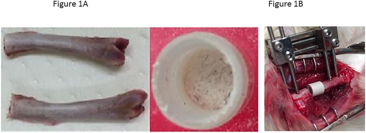
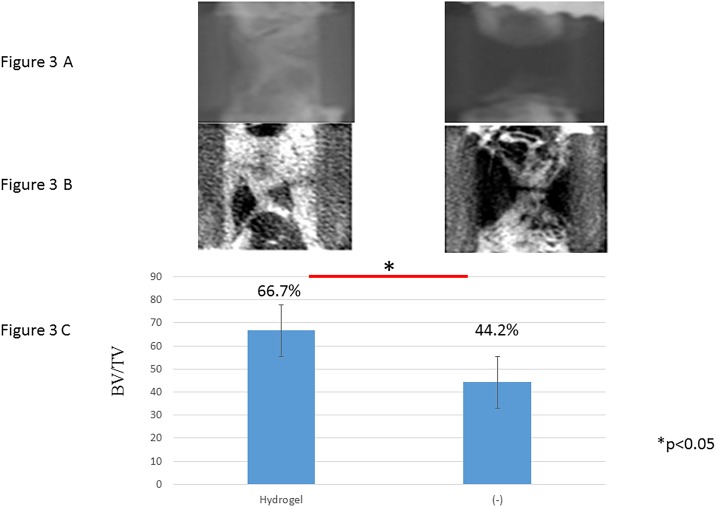
Spinal fusion and bone defect after injuries, removal of bone tumors, and infections need to be repaired by implantation. In an aging society, recovery from these procedures is often difficult. In this study, we found that injection of SPG-178 leads to expression of several bone marker genes and mineralization in vitro, and revealed a significantly higher degree of newly formed bone matrix with use of SPG-178 in vivo. MC3T3-E1 cells were used to evaluate osteoblast differentiation promoted by SPG-178. To analyze gene expression, total RNA was isolated from MC3T3-E1 cells cultured for 7 and 14 days with control medium or SPG-178 medium. Among the several bone marker genes examined, SPG-178 significantly increased the mRNA levels for ALP, BMP-2 and Osteocalcin, OPN, BSP and for the Osterix. Ten-week-old female Wistar rats were used for all transplantation procedures. A PEEK cage was implanted into a bony defect (5 mm) within the left femoral mid-shaft, and stability was maintained by an external fixator. The PEEK cages were filled with either a SPG-178 hydrogel plus allogeneic bone chips (n = 4) or only allogeneic bone chips (n = 4). The rats were then kept for 56 days. Newly formed bone matrix was revealed inside the PEEK cage and there was an increased bone volume per total volume with the cage filled with SPG-178, compared to the control group. SPG-178 has potential in clinical applications because it has several benefits. These include its favorable bone conduction properties its ability to act as a support for various different cells and growth factors, its lack of infection risk compared with materials of animal origin such as ECM, and the ease with which it can be used to fill defects with complex shapes and combined with a wide range of other materials.
Conflict of interest statement
Figures


References
-
- Feuvrier D, Sagawa Y Jr., Beliard S, Pauchot J, Decavel P. Long-term donor-site morbidity after vascularized free fibula flap harvesting: Clinical and gait analysis. Journal of plastic, reconstructive & aesthetic surgery: JPRAS. 2016;69(2):262–9. doi: 10.1016/j.bjps.2015.10.007 . - DOI - PubMed
-
- Lee SS, Huang BJ, Kaltz SR, Sur S, Newcomb CJ, Stock SR, et al. Bone regeneration with low dose BMP-2 amplified by biomimetic supramolecular nanofibers within collagen scaffolds. Biomaterials. 2013;34(2):452–9. doi: 10.1016/j.biomaterials.2012.10.005 - DOI - PMC - PubMed
-
- Shang Q, Wang Z, Liu W, Shi Y, Cui L, Cao Y. Tissue-engineered bone repair of sheep cranial defects with autologous bone marrow stromal cells. The Journal of craniofacial surgery. 2001;12(6):586–93; discussion 94–5. . - PubMed
-
- Parikh SN. Bone graft substitutes: past, present, future. Journal of postgraduate medicine. 2002;48(2):142–8. . - PubMed
-
- Sengupta S, Park SH, Patel A, Carn J, Lee K, Kaplan DL. Hypoxia and amino acid supplementation synergistically promote the osteogenesis of human mesenchymal stem cells on silk protein scaffolds. Tissue engineering Part A. 2010;16(12):3623–34. doi: 10.1089/ten.TEA.2010.0302 - DOI - PMC - PubMed
Publication types
MeSH terms
Substances
LinkOut - more resources
Full Text Sources
Other Literature Sources
Research Materials
