. 2018 Feb 1;77(2):149-161.
doi: 10.1093/jnen/nlx113.

Selective Vulnerability of Brainstem Nuclei in Distinct Tauopathies: A Postmortem Study

Affiliations

Selective Vulnerability of Brainstem Nuclei in Distinct Tauopathies: A Postmortem Study

Rana A Eser et al. J Neuropathol Exp Neurol. .

Abstract

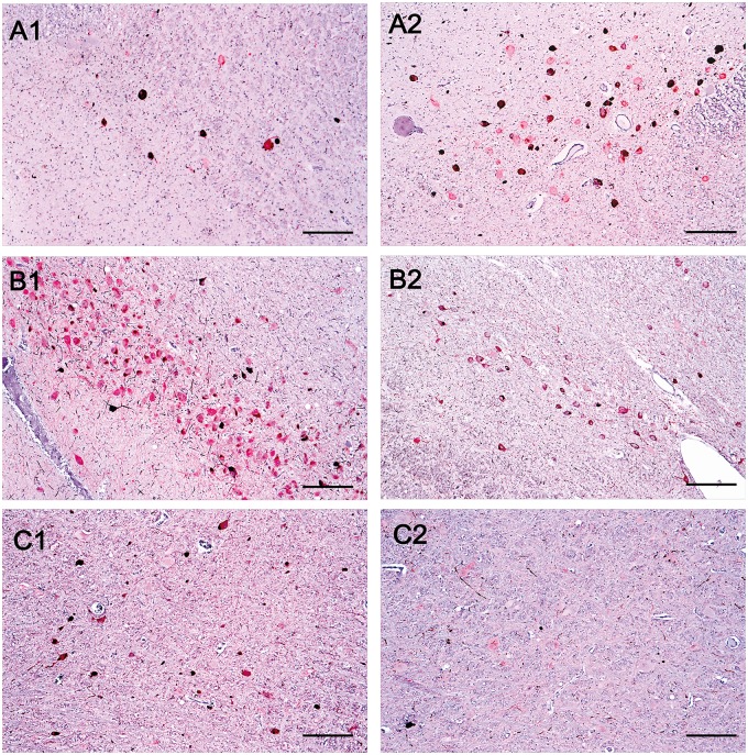
The brainstem nuclei of the reticular formation (RF) are critical for regulating homeostasis, behavior, and cognition. RF degenerates in tauopathies including Alzheimer disease (AD), progressive supranuclear palsy (PSP), and corticobasal degeneration (CBD). Although the burden of phopho-tau inclusion is high across these diseases, suggesting a similar vulnerability pattern, a distinct RF-associated clinical phenotype in these diseases indicates the opposite. To compare patterns of RF selective vulnerability to tauopathies, we analyzed 5 RF nuclei in tissue from 14 AD, 14 CBD, 10 PSP, and 3 control cases. Multidimensional quantitative analysis unraveled discernable differences on how these nuclei are vulnerable to AD, CBD, and PSP. For instance, PSP and CBD accrued more tau inclusions than AD in locus coeruleus, suggesting a lower vulnerability to AD. However, locus coeruleus neuronal loss in AD was so extreme that few neurons remained to develop aggregates. Likewise, tau burden in gigantocellular nucleus was low in AD and high in PSP, but few GABAergic neurons were present in AD. This challenges the hypothesis that gigantocellular nucleus neuronal loss underlies REM behavioral disorders because REM behavioral disorders rarely manifests in AD. This study provides foundation for characterizing the clinical consequences of RF degeneration in tauopathies and guiding customized treatment.

Keywords: Alzheimer disease; Corticobasal degeneration; Human brainstem; Progressive supranuclear palsy; Reticular formation; Selective vulnerability; Tauopathies.


Figures

FIGURE 1.
FIGURE 1.
A diagram illustrating how cells were classified for analysis. Cells were classified as positive for the neurotransmitter of interest (NEU+) as indicated by the color red, or negative for the neurotransmitter of interest (NEU+) as indicated by a colorless circle. Cells were also classified by the presence of tau inclusions (tau+) indicated by a brown coloring within the cell, or the absence of tau inclusions (tau+). This generated 4 different groups: (i) number of colocalized neurons, (ii) number of NEU+ neurons, (iii) number of tau+ neurons, and (iv) the number of total neurons, including those negative for both tau and the neurotransmitter.
FIGURE 2.
FIGURE 2.
Histological section (8-µm-thick) through the (A) dorsal raphe nucleus (double immunostained for phospho-tau [CP-13, in brown] and tryptophan hydroxylase [PH8, in red]) and (B) substantia nigra (CP-13, in brown) and tyrosine hydroxylase (PH8, in red)). The figure depicts some examples on how the neurons were classified: (1 and 6) tau-positive/NEU-positive colocalized; (2 and 5) neuron positive for neurotransmitter only (NEU-positive); (3) positive for phospho-tau only (tau-positive); and (4) neuron negative for phospho-tau and not expressing the neurotransmitter of interest.
FIGURE 3.
FIGURE 3.
A multidimensional visual representation of the data of each of the 5 nuclei: locus coeruleus (A), substantia nigra (B), gigantocellular nucleus (C), pedunculopontine nucleus (D), dorsal raphe nucleus (E). The x-axis is the proportion of neurons bearing tau (tau+) and the y-axis is the proportion of neurons that express the neurotransmitter of interest (NEU+). The size of each dot is directly proportional to the neuronal numbers. The color of the dot indicates the diagnosis: Alzheimer disease (AD), progressive supranuclear palsy (PSP), corticobasal degeneration (CBD), or control.
FIGURE 4.
FIGURE 4.
Neuroanatomical schemes showing the location of each nucleus of interest and the degree of difference between the disease compared with the control for the variable. The figure depicts the total number of neurons (upper row), percent of tau-bearing (tau-positive) neurons (middle row), and percent of neurotransmitter-synthesizing (NEU-positive) neurons (bottom row).
FIGURE 5.
FIGURE 5.
Comparison of histopathological features between Alzheimer disease (AD), corticobasal degeneration (CBD) and progressive supranuclear palsy (PSP). (A) Histological slides containing the locus coeruleus immunostained for phospho-tau and TH of an AD case (A1) and in a PSP case (A2). (B) Histological slides containing the substantia nigra immunostained for phospho-tau and TH of an AD case (B1) and a CBD case (B2). (C) Histological slides containing the gigantocellular nucleus in a PSP case (C1) and an AD case (C2). Scale bar: 25 µm.

References

    1. Vogt O, Vogt C.. Erkrankungen Der Grosshirnrinde Im Lichte Der Topistik, Pathoklise Und Pathoarchitektonik. Leipzig: J.A. Barth; 1922
    1. Saxena S, Caroni P.. Selective neuronal vulnerability in neurodegenerative diseases: From stressor thresholds to degeneration. Neuron 2011;71:35–48http://dx.doi.org/10.1016/j.neuron.2011.06.031 - DOI - PubMed
    1. Theofilas P, Dunlop S, Heinsen H, et al.Turning on the light within: Subcortical nuclei of the isodentritic core and their role in Alzheimer’s disease pathogenesis. J Alzheimers Dis 2015;46:17–34 - PMC - PubMed
    1. Ramon-Moliner E, Nauta WJ.. The isodendritic core of the brain stem. J Comp Neurol 1966;126:311–35http://dx.doi.org/10.1002/cne.901260301 - DOI - PubMed
    1. Nieuwenhuys R. The Human Central Nervous System. 4th ed.New York: Springer Berlin Heidelberg; 2008

Publication types