. 2018 Jan 5;11(1):4.
doi: 10.1186/s13048-017-0376-6.

A protocol to isolate and qualify purified human preantral follicles in cases of acute leukemia, for future clinical applications

Affiliations

A protocol to isolate and qualify purified human preantral follicles in cases of acute leukemia, for future clinical applications

Elodie Mouloungui et al. J Ovarian Res. .

Abstract

Background: Autotransplantation of cryopreserved ovarian cortex can be associated with a risk of cancer cell reseeding. This issue could be eliminated by grafting isolated preantral follicles. Collagenase NB6 is an enzyme produced under good manufacturing practices (GMP) in compliance with requirements for tissue engineering and transplantation in humans and thus can be used to isolate preantral follicles from ovarian tissue in the framework of further clinical applications. Multicolor flow cytometry is an effective tool to evaluate the potential contamination of follicular suspensions by leukemic cells.

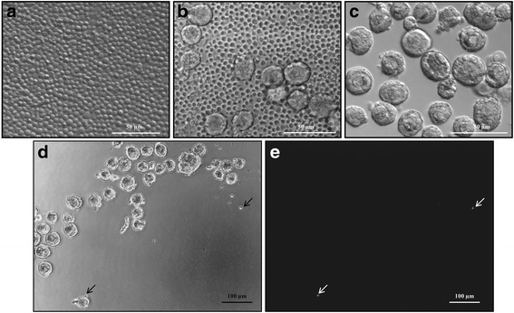
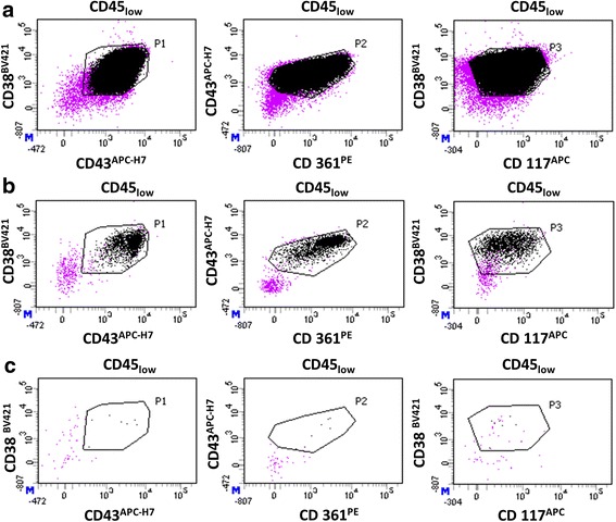
Methods: The efficiency of collagenase NB6 was evaluated in comparison to collagenase type IA and Liberase DH, in terms of yield, morphology and viability. A short-term in vitro culture of follicles isolated with collagenase NB6 was conducted for 3 days in a fibrin matrix. A modelization procedure was carried out to detect the presence of leukemic cells in follicular suspensions using multicolor flow cytometry (MFC).

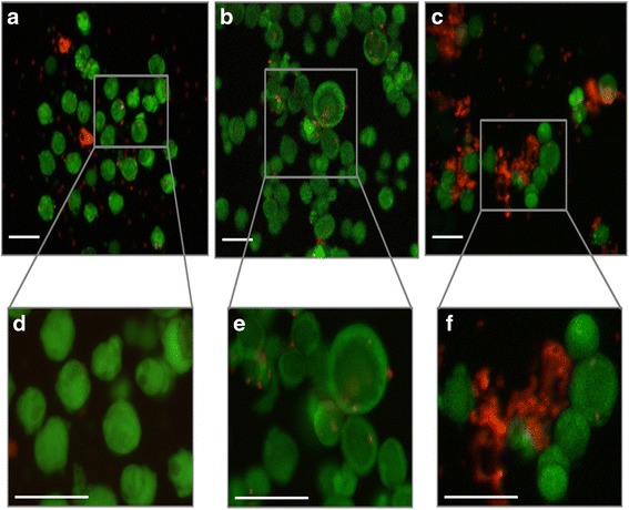
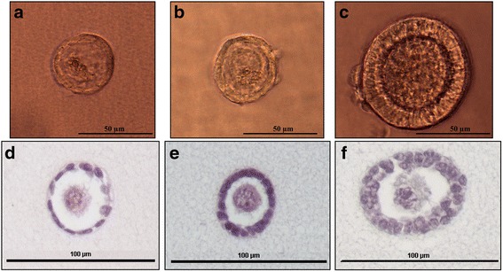
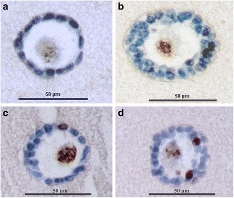
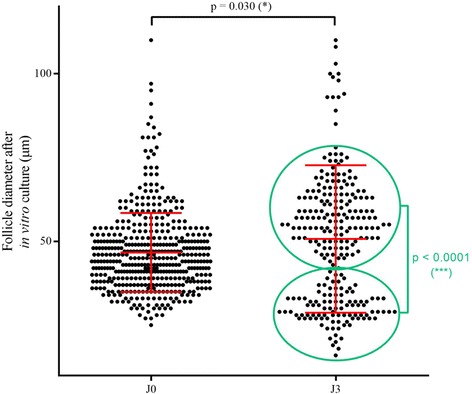
Results: No statistical differences were found between collagenase NB6, Liberase DH (p = 0.386) and collagenase type IA (p = 0.171) regarding the number of human preantral follicles isolated. The mean diameter of isolated follicles was significantly lower with collagenase NB6 (p < 0.0001). The survival rate of isolated follicles was 93.4% (n = 272) using collagenase NB6 versus 94.9% (n = 198) with Liberase DH and 92.6% (n = 298) using collagenase type IA. Even after 3 days of in vitro culture in a fibrin scaffold, most of the isolated follicles were still alive after using collagenase NB6 (90.7% of viable follicles; n = 339). The rate of isolated Ki67-positive follicles was 29 ± 9.19% before culture and 45 ± 1.41% after 3 days. In 23 out of 24 follicular suspensions analyzed, the detection of leukemic cells by MFC was negative. The purification had no significant impact on follicle viability.

Conclusion: The isolation and purification of human preantral follicles were performed following good manufacturing practices for cell therapy. Multicolor flow cytometry was able to confirm that final follicular suspensions were free from leukemic cells. This safe isolation technique using collagenase NB6 can be considered for future clinical applications.

Keywords: Collagenase NB6; Good manufacturing practices; Human follicle isolation; Leukemic cell purification.


Conflict of interest statement

Ethical approval and consent to participate

The use of human ovarian tissue was approved by the clinical ethics committee of Besancon University Hospital in 2013; all patients gave their informed consent. The use of leukemic cells from leukemia patients was approved by the department of research and innovation (CRB F.Cabanne, Dijon-Besançon, France; biological collection authorization n°DC-2008-713). The use of AB human serum from male donors was approved by the French blood establishment (request of blood samples for non-therapeutic uses, BFC/PSL/COL/FO/014).

Consent for publication

Not applicable.

Competing interests

The authors declare that they have no competing interests.

Publisher’s Note

Springer Nature remains neutral with regard to jurisdictional claims in published maps and institutional affiliations.

Figures

Fig. 1
Fig. 1
Viability assay performed on isolated follicles using the fluorescent probes calcein AM and ethidium homodimer-1. a and d. Isolated follicles after using collagenase NB6; b and e. Isolated follicles after using Liberase DH; c and f. Isolated follicles after using collagenase IA; green: live follicles; red: dead follicles; scale bar = 100 μm
Fig. 2
Fig. 2
Isolated preantral follicles embedded in a fibrin scaffold. Brightfield images of an isolated primordial (a), primary (b) and secondary (c) follicle inside a fibrin scaffold; Hematoxylin and eosin staining of an isolated primordial (d), primary (e) and secondary (f) follicle inside a fibrin scaffold
Fig. 3
Fig. 3
Immunohistochemistry of isolated preantral follicles embedded in a fibrin scaffold. a Embedded isolated primordial follicle with Ki67-negative granulosa cells in blue; b Embedded isolated secondary follicle with Ki67-negative granulosa cells in blue; c Embedded isolated primary follicles with one Ki67-positive granulosa cell in brown; d Embedded isolated secondary follicles with two Ki67-positive granulosa cells in brown
Fig. 4
Fig. 4
Follicle diameter before and after 3 days of short-term in vitro culture in a fibrin gel. Dots represent each embedded follicles measured. * = statistically significant
Fig. 5
Fig. 5
Cell suspensions observed under an inverted micromanipulation microscope. a Leukemic cell suspension obtained after thawing, corresponding to positive controls; b Ovarian cell suspension, containing follicles and leukemic cells, without wash; c Ovarian cell suspension obtained after 3 washes of isolated follicles; d An example of ovarian cell suspension obtained after 3 washes of isolated follicles, with two leukemic cells visible (black arrows); e The same ovarian cell suspension obtained after 3 washes under inverted fluorescence microscope, showing the presence of two leukemic cells stained by using a fluorescent cell tracer (white arrows)
Fig. 6
Fig. 6
Quantification of leukemic cells by MFC in an ovarian suspension containing isolated follicles. Only viable nucleated cells (SYTO13+/7-AAD), which are CD45low were represented (purple dots). Black dots represent leukemic cells detected with a typical immunophenotype CD38+/CD43+/CD361+/CD117+ among the population of CD45low cells. A positive event must be at the intersection of gates P1 (CD38 versus CD43), P2 (CD43 versus CD361), and P3 (CD38 versus CD117). a Positive control (leukemic cells); b MFC analysis of follicles isolated from healthy ovarian suspension after addition of 106 leukemic cells, before any wash (n = 2069 events detected); c MFC analysis of isolated follicles after 3 washes (n = 7 events detected)

Similar articles

Cited by

References

    1. Siegel R, Ma J, Zou Z, Jemal A. Cancer statistics, 2014. CA Cancer J Clin. 2014;64(1):9–29. doi: 10.3322/caac.21208. - DOI - PubMed
    1. Ward E, DeSantis C, Robbins A, Kohler B, Jemal A. Childhood and adolescent cancer statistics, 2014. CA Cancer J Clin. 2014;64(2):83–103. doi: 10.3322/caac.21219. - DOI - PubMed
    1. Imbert R, Moffa F, Tsepelidis S, Simon P, Delbaere A, Devreker F, et al. Safety and usefulness of cryopreservation of ovarian tissue to preserve fertility: a 12-year retrospective analysis. Hum Reprod. 2014;29(9):1931–1940. doi: 10.1093/humrep/deu158. - DOI - PubMed
    1. Lee SJ, Schover LR, Partridge AH, Patrizio P, Wallace WH, Hagerty K, et al. American Society of Clinical Oncology recommendations on fertility preservation in cancer patients. J Clin Oncol. 2006;24(18):2917–2931. doi: 10.1200/JCO.2006.06.5888. - DOI - PubMed
    1. Peddie VL, Porter MA, Barbour R, Culligan D, MacDonald G, King D, et al. Factors affecting decision making about fertility preservation after cancer diagnosis: a qualitative study. BJOG. 2012;119(9):1049–1057. doi: 10.1111/j.1471-0528.2012.03368.x. - DOI - PubMed